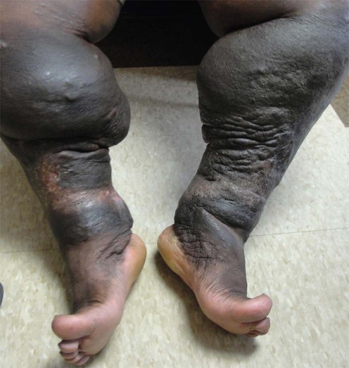
138 Lymphedema Robert E. Lee, Giuseppe Micali and Robert A. Schwartz Evidence Levels: A Double-blind study B Clinical trial ≥ 20 subjects C Clinical trial < 20 subjects D Series ≥ 5 subjects E Anecdotal case reports Lymphedema is a chronic, progressive, and sometimes debilitating condition. It is due to the abnormal accumulation of protein-rich lymphatic fluid in interstitial spaces as a result of ineffective drainage. Lymphedema is classified into primary and secondary forms. Primary lymphedema is caused by a developmental malformation of the lymphatic system. Primary congenital lymphedema (Milroy disease) is an uncommon autosomal dominant disorder due, in some families, to missense mutations that interfere with vascular endothelial growth factor receptor-3 signaling, resulting in abnormal lymphatic vascular function. Primary lymphedema may be further classified by age of onset into congenital lymphedema (before age 2 years), lymphedema praecox (between age 2 and 35 years), and lymphedema tarda (after age 35 years). Secondary lymphedema is due to obstruction or damage of an otherwise normal lymphatic system. In the United States the most common causes are malignancy, surgical manipulation, or radiation damage. Globally, the most common cause is filariasis. Acquired lymphedema may predispose to recurrent cellulitis and an aggressive type of angiosarcoma, also known as Stewart–Treves syndrome. Chronic lymphedema may result in verrucous and proliferative changes resembling elephant skin (elephantiasis). Management strategies Lymphedema must be distinguished from cutaneous edema of cardiac, hepatic, or renal etiology. It is characterized clinically by brawny, non-pitting edema. Lymphoscintigraphy (isotope lymphography) is a first-line imaging modality to evaluate and diagnose disorders of the lymphatic vasculature. A conservative approach with medical and physical therapy is the primary treatment for lymphedema. The main management strategy is to reduce stasis of protein-rich lymph in the extravascular tissue and to improve the outflow of lymphatic circulation. Complex decongestive therapy (CDT) represents an effective treatment plan. It is a four-component therapeutic modality composed of multilayer compression bandaging, manual lymphatic drainage, skin care, and exercise. The therapeutic efficacy of CDT is highly dependent upon patient compliance. Therefore, strict adherence to the treatment regimen should be encouraged. Pneumatic compression therapy was widely used to control lymphedema. However, owing to its poor outcome as a monotherapy, its current use has been limited. Unfortunately, recent advances in these devices have showed beneficial results. Medications, such as diuretics, have shown limited or no effect on lymphedema. Meticulous skin care and hygiene may prevent secondary bacterial and fungal infections. Topical or systemic antibiotics should be initiated at the first sign of infection. This is especially important as recurrent infections may lead to further lymphatic injury. Surgical approaches are reserved for cases refractory to conservative medical management. Microsurgical lymphatic–venous anastomoses have yielded promising results. Excisional surgical therapy has been performed to reduce limb size and to improve mobility in chronic advanced cases of lymphedema. Specific investigations Lymphoscintigraphy MRI CT Indocyanine green lymphography Only gold members can continue reading. Log In or Register to continue Related Related posts: Cat scratch disease Mucoceles Tinea capitis Herpes genitalis Necrolytic migratory erythema Nevoid basal cell carcinoma syndrome Stay updated, free articles. Join our Telegram channel Join Tags: Treatment of Skin Disease Comprehensive Therapeutic Strategies Aug 7, 2016 | Posted by admin in Dermatology | Comments Off on Lymphedema Full access? Get Clinical Tree
138 Lymphedema Robert E. Lee, Giuseppe Micali and Robert A. Schwartz Evidence Levels: A Double-blind study B Clinical trial ≥ 20 subjects C Clinical trial < 20 subjects D Series ≥ 5 subjects E Anecdotal case reports Lymphedema is a chronic, progressive, and sometimes debilitating condition. It is due to the abnormal accumulation of protein-rich lymphatic fluid in interstitial spaces as a result of ineffective drainage. Lymphedema is classified into primary and secondary forms. Primary lymphedema is caused by a developmental malformation of the lymphatic system. Primary congenital lymphedema (Milroy disease) is an uncommon autosomal dominant disorder due, in some families, to missense mutations that interfere with vascular endothelial growth factor receptor-3 signaling, resulting in abnormal lymphatic vascular function. Primary lymphedema may be further classified by age of onset into congenital lymphedema (before age 2 years), lymphedema praecox (between age 2 and 35 years), and lymphedema tarda (after age 35 years). Secondary lymphedema is due to obstruction or damage of an otherwise normal lymphatic system. In the United States the most common causes are malignancy, surgical manipulation, or radiation damage. Globally, the most common cause is filariasis. Acquired lymphedema may predispose to recurrent cellulitis and an aggressive type of angiosarcoma, also known as Stewart–Treves syndrome. Chronic lymphedema may result in verrucous and proliferative changes resembling elephant skin (elephantiasis). Management strategies Lymphedema must be distinguished from cutaneous edema of cardiac, hepatic, or renal etiology. It is characterized clinically by brawny, non-pitting edema. Lymphoscintigraphy (isotope lymphography) is a first-line imaging modality to evaluate and diagnose disorders of the lymphatic vasculature. A conservative approach with medical and physical therapy is the primary treatment for lymphedema. The main management strategy is to reduce stasis of protein-rich lymph in the extravascular tissue and to improve the outflow of lymphatic circulation. Complex decongestive therapy (CDT) represents an effective treatment plan. It is a four-component therapeutic modality composed of multilayer compression bandaging, manual lymphatic drainage, skin care, and exercise. The therapeutic efficacy of CDT is highly dependent upon patient compliance. Therefore, strict adherence to the treatment regimen should be encouraged. Pneumatic compression therapy was widely used to control lymphedema. However, owing to its poor outcome as a monotherapy, its current use has been limited. Unfortunately, recent advances in these devices have showed beneficial results. Medications, such as diuretics, have shown limited or no effect on lymphedema. Meticulous skin care and hygiene may prevent secondary bacterial and fungal infections. Topical or systemic antibiotics should be initiated at the first sign of infection. This is especially important as recurrent infections may lead to further lymphatic injury. Surgical approaches are reserved for cases refractory to conservative medical management. Microsurgical lymphatic–venous anastomoses have yielded promising results. Excisional surgical therapy has been performed to reduce limb size and to improve mobility in chronic advanced cases of lymphedema. Specific investigations Lymphoscintigraphy MRI CT Indocyanine green lymphography Only gold members can continue reading. Log In or Register to continue Related Related posts: Cat scratch disease Mucoceles Tinea capitis Herpes genitalis Necrolytic migratory erythema Nevoid basal cell carcinoma syndrome Stay updated, free articles. Join our Telegram channel Join Tags: Treatment of Skin Disease Comprehensive Therapeutic Strategies Aug 7, 2016 | Posted by admin in Dermatology | Comments Off on Lymphedema Full access? Get Clinical Tree