Lower extremity sarcoma reconstruction

4 Lower extremity sarcoma reconstruction


Introduction


Soft-tissue and bone sarcomas


Soft-tissue tumors are a highly heterogeneous group of about 100 different tumor entities that are classified histogenetically according to their main adult tissue component they resemble most. The malignant subgroup of soft-tissue tumors is called sarcomas, which not only have the potential to grow locally invasive or even demonstrate destructive growth, but also have a variable risk of recurrence and metastatic potential. As the term “sarcoma” (derived from the Greek word σαρξ, sarx = meat) does not necessarily imply fast, expansive growth or metastasis, a further subclassification system into more aggressive sarcomas (high-grade, poorly differentiated) or less aggressive (low-grade, well-differentiated) types exists. Some lesions, like the atypical fibroxanthoma, are called “pseudosarcomas,” as they demonstrate a benign clinical course but are histologically malignant. However, well-differentiated tumors usually show low-grade characteristics and vice versa.


Primary bone sarcomas are even less frequent than soft-tissue sarcomas, and most malignant osseous lesions are metastatic, especially in advanced age. Despite having a low incidence, bone sarcomas have a high significance for both patient and surgeon due to their impact on extremity function and overall mobility. Limb-preserving surgery after wide tumor resections frequently poses a real challenge for the reconstructive surgeon.


Sarcomas can occur in every part of the body, as they derive from mesodermic tissue like muscle, nerve, bone cartilage, blood vessels, or fat. The therapeutic mainstay for soft-tissue sarcomas is based on surgical excision whereas radiotherapy and, less so, chemotherapy are used as adjunct adjuvant therapeutic modalities. In primary osseous sarcomas neoadjuvant chemotherapy plays a more important role. The complexity of the surgical resection and the subsequent plastic surgical reconstruction differs considerably depending on the localization.


Like any oncologic discipline, sarcoma treatment is a typical field of modern interdisciplinary and multimodal therapy. Lower extremity sarcoma reconstruction, especially in bone sarcoma reconstruction, is also a fascinating field for an effective interaction between the surgical disciplines of oncologic, pediatric and podiatric surgeons, orthopedic surgeons and plastic reconstructive (micro-) surgeons.


Modern plastic reconstructive surgery can provide adequate reconstructive options for almost any defect size and composition, so considerations about defect size should not play a role in tumor excision. Over 95% of the extremities can be preserved after radical tumor excision today. The plastic reconstructive procedures are often demanding and complex and often encompass the full spectrum of plastic surgical options. These procedures may be best performed in specialized centers where individually adapted regimens to patient and case profiles may be optimized.


Sarcomas in the lower extremity


Sarcomas in the lower extremity are more common than in the upper limb (74 versus 26%) and represent the most common location of sarcomas in the body overall (45%).1 Currently, they can be safely treated by extremity preservation in most cases, if properly performed according to the rules described in this chapter and in the pertinent current literature. In this context, several studies have now demonstrated that limb-sparing surgery is oncologically not inferior to amputation in the treatment of lower extremity sarcoma (see Outcomes, prognosis, and complications, below). While amputation was the keystone of previous surgical therapy several decades ago, it usually represents only an important last-line therapeutic modality today. The common misconceptions that amputations have a better outcome in both tumor safety and quality of life have both definitely been proven wrong.24


Sarcomas are rare and a “soft-tissue swelling” is often misinterpreted by both patient and physician because of this fact. This can considerably delay proper diagnosis. Still, tumor manifestations in the extremities are often detected slightly earlier than in the trunk, as the extremities are constantly under personal “visual” control in daily life. This may be even truer for tumors on the upper extremity than on the lower extremity.


Due to the highly functional anatomy of our extremities with vessels, nerves, tendons, bones, and muscles in close vicinity, even smaller tumors can represent a challenge to both the resecting tumor surgeon as well as the reconstructive plastic surgeon. Preservation of the lower extremity in sarcoma reconstruction differs from similar manifestations in the upper extremity5 in several key points:


Basic science/disease process


Epidemiology soft-tissue sarcomas


Soft-tissue sarcomas are a rare disease entity with an incidence of 1 : 100 000 in adults and 10–15% in children. This accounts for an incidence of about 10 600 new cases in 2009 in the US, representing 1–2% of all malignancies (www.seer.cancer.gov). There is no overall significant gender predisposition and the overall median age at presentation is 50–60 years.


With about 45% of all sarcomas occurring in the lower extremity, 15% in the upper extremity, 10% in the head and neck region, 15% in the retroperitoneal space, and the remaining 15% in the abdomen and the chest wall,6 the musculoskeletal system of the extremities and the abdominal and thoracic walls is the most common predilection site. Extremity sarcomas are most common in the thigh (50–60%).


While most cases of soft-tissue sarcomas are sporadic, there are some genetic and nongenetic risk factors, summarized in Table 4.1. Up to 60% of all soft-tissue sarcomas contain a somatic mutation of p53.7 A detailed description of the various risk factors is beyond the scope of this chapter, but there are several strong associations to be mentioned: a history of radiation exposure accounts for up to 5.5% of all sarcomas. The risk is dose-dependent and the latency period between radiation and clinical tumor manifestation is around 5 years. Over 80% of radiation-associated sarcomas are high-grade types.8 Neurofibromatosis type NF-1 is strongly associated with the cumulative lifetime risk of up to 13% for the occurrence of malignant peripheral nerve sheath tumors.


Table 4.1 Predisposing factors for soft-tissue sarcomas


Genetic Neurofibromatosis NF-1 (von Recklinghausen disease)
  Retinoblastoma
  Gardner’s syndrome
  Werner’s syndrome
  Bloom’s syndrome
  Fumarate hydratase leiomyosarcoma syndrome
  Diamond–Blackfan anemia
Mechanical Li–Fraumeni syndrome
  Postparturition
Chemical Chronic irritation
  Polyvinylchloride
  Hemochromatosis
  Dioxin (TCDD): “Agent Orange”
Radiation Arsenic
  Traumatic/accidental
Lymphedema Posttherapeutic
  Parasitic (filariasis)
  Iatrogenic
  Stewart–Treves syndrome
Infectious (viral) Congenital
  Kaposi sarcoma (human herpesvirus-8)

The sarcoma subtype is determined by light and electron microscopy, immunohistochemistry, and cytogenetic analysis. If it results in a tumor that cannot be designated accordingly, a descriptive evaluation is given for an “unclassified sarcoma.” Obtaining reference pathologies for soft- and bone tissue sarcomas should be standard, as the rate of diagnostic agreement among specialists is below 75%.9,10


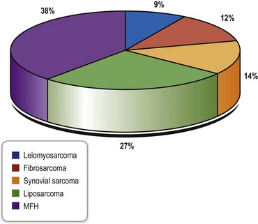
The most common histopathologic subtype distribution in extremities in the largest series in the literature is shown in Figure 4.1.


Bone sarcomas


About 2600 new primary bone sarcomas occur each year in the US (www.seer.cancer.gov). The overall median age at diagnosis is 39 years. Many predisposing factors for bone sarcomas are similar to those for soft-tissue sarcomas (retinoblastoma, Li–Fraumeni syndrome, radiation, and others) (Table 4.1). Paget’s disease, bone infarction, and fibrous dysplasia may also represent risk factors for bone sarcomas.


The most common type is the osteogenic sarcoma, which has a predilection for the metaphyses around the knee in about 50% of cases. It is the third most common cancer in the young (www.nhs.uk) with a second peak around age 60. The male-to-female ratio is almost 2 : 1 in large studies and for this specific tumor the median age at diagnosis is 17 years. Only 6.4% present initially with pathological fractures, whereas the majority are detected in the workup of a painful mass or swollen extremity.11,12 It commonly arises in the medulla, but as a juxtacortical osteogenic sarcoma it arises from the external surface, most commonly the posterior aspect of the femur.


The spindle cell mesenchymal sarcoma group contains chondrosarcomas, intraosseous malignant fibrous histiocytomas, and fibrosarcomas. The tumors of this group only have an incidence of about two-thirds of that of osteogenic sarcomas and primarily occur in an older population. Chondrosarcomas are slow-growing and relatively resistant to adjuvant therapy.


Ewing’s sarcoma is classically located in the femur diaphyses in teenagers and only 20% occur in middle-aged adults. If extradiaphyseal, it is very common in the pelvis. It is the most common primary bone malignancy of the fibula. Ewing’s sarcoma is very radiation-sensitive.


Historical perspective


The term “sarcoma” was first used by Abernethy in 1804, based on gross characteristic of the tumors. Codman founded the first Bone and Soft Tissue Sarcoma Registry in 1909, containing information on the diagnosis and treatment of bone. In 1829 Jean Cruveilhier published a two-volume work on pathologic anatomy, containing a substantial amount of information about sarcomas as they are understood today.


Since then, the classical treatment of a sarcoma in the lower extremity was amputation, accounting for relatively low recurrence rates, but with a severe impact on the integrity of the patient. In 1879, Samuel W Gross published his experiences with 165 sarcoma cases of the long bones, in which he advocated early amputation despite the prevailing operative mortality rate of 30%. Based on his study, limb-salvaging resection inevitably led to local recurrence, metastasis, and death. Following this study, an even more aggressive approach to bone tumors was popularized, but survival rates did not improve significantly. This stimulated the first adjuvant radiation treatment of bone tumors.


However, the mortality rate of radiated patients equaled the mortality rate of patients who underwent amputation. A first neoadjuvant protocol was developed in 1940 by Cade and Ferguson,15 who used preoperative radiation followed by amputation 6 months later in metastasis-free patients. The aim of the protocol was to avoid unnecessary amputation.


The transition between amputation surgery and limb-sparing surgery was less of a plastic surgical issue than an oncologic–orthopedic development: reconstructive procedures after sarcoma resection were very uncommon until the number of limb-preserving resections increased.


Episodic anecdotes reporting limb salvage appear as early as 1895, when Mikulicz described two resection arthrodeses of the knee for distal femoral lesions in Europe.16 Sauerbruch in Germany described his “Umkippplastik” in 1922 as the precursor of today’s rotationplasty.17 A first systematic approach to limb salvage was suggested in 1940 by Phemister in his article “Conservative surgery in the treatment of bone tumors”.18,19


After World War II more surgeons began to explore limb-salvaging resection instead of amputation. It became clear that obtaining adequate surgical margins is a key issue for cases with resectable tumors according to their pathologic characteristics. Their work established the principles of today’s limb-salvaging surgery despite the limited reconstructive modalities at that time.


The surgical treatment of malignant bone tumors was revolutionized in the early 1970s by the development of chemotherapy, improved diagnostic radiology with more accurate computed tomography (CT) and magnetic resonance imaging (MRI) scanners, advances in reconstructive surgery, improvements in orthopedic oncologic surgery, and eventually the establishment of multidisciplinary tumor centers. These advances are mainly responsible for the reduction in local recurrences after limb-salvaging procedures by allowing better patient selection and more accurate preoperative planning.


During these developments, many sophisticated plastic surgical techniques were developed in a fascinating history which is covered in a different volume of this textbook. Most of the techniques focused on soft-tissue reconstruction, but later composite tissue transplants and grafts were improved as well. Many new techniques derived from trauma surgery, the treatment of congenital disorders or tumor surgery elsewhere in the body and did not specifically relate to the development of sarcoma surgery, but were quickly adapted to limb-preserving tumor reconstruction. Sophisticated pedicled reconstructive methods like cross-leg flaps, tubed pedicled flaps, or fibula-pro-tibia transfers were standard procedures in early limb-sparing surgery until microvascular surgery was integrated into the plastic surgical armamentarium.


Modern reconstructive approaches integrate the latest advances of orthopedic surgery like modular and custom-made tumor prostheses for long bone and joint replacement with the full spectrum of the plastic surgical reconstructive ladder, including chimeric multitissue-type free flaps in limb-sparing lower extremity sarcoma reconstruction.


Diagnosis/patient presentation/imaging


A detailed history and physical examination are the first initial and important steps to professional tumor surgery. In sarcomatous lesions, the patient often relates the tumor causally to an – often minor – traumatic event that brings the lesion to the patient’s attention. Acute trauma, however, is not a proven predisposing factor for sarcoma development. Because of this, there is often a considerable time lag between this initial recognition and the first presentation of the lesion to a medical professional. Furthermore, the rationale of the lesion is often erratically misinterpreted and then causes a variety of inadequate treatments by both lays and physicians, further delaying proper diagnosis. The average duration of any symptoms before seeing a physician is 6 months in all soft-tissue sarcomas, but possibly shorter in extremity manifestation.3 So in adults, lesions that: (1) have not disappeared after 4 weeks; (2) are located subfascially or in the popliteal or groin flexion creases; (3) continue to grow or are symptomatic (i.e., pain, paresthesias); or (4) are already larger than 5 cm on detection should generally be biopsied as they are highly suspicious for malignancy.


It is not unusual that sarcomas are found by physicians in the context of workup of a completely different medical problem (i.e., chronic venous insufficiency in the leg). Coincidental findings like articular pain and joint effusions are common, especially with osseous sarcomas, whereas clinically manifest neurovascular symptoms are relatively rare at initial presentation. Two-thirds of sarcoma patients present with a painless mass during their first clinical examination, and only one-third have current pain or have had a history of pain in the affected region.


The thorough physical examination is not only focused on the affected extremity but includes the complete body. The pertaining lymph node stations should be examined as well, even though lymphatic spread is uncommon in the majority of all sarcoma types. The general health status should be assessed and optimized by all relevant medical specialties. This is especially important in multimorbid patients with concomitant acute and chronic comorbidities in the context of planned operative procedures. Terminal illnesses and comorbidities have to be taken into consideration for the extent of both resection and reconstructive surgery.


Clinical assessment and staging of the patient must be completed by adequate imaging diagnostics for evaluating local and generalized tumor manifestations. Any imaging of the tumor region must be performed before surgical biopsy as the latter may confound the picture to a considerable extent.


Gadolinium contrast-enhanced MRI is currently the diagnostic mainstay to define exact tumor location, its relation to neighboring neurovascular structures and muscular compartments, to determine its homogeneity, integrity, vascularization, and its presumed main tissue component. MRI is specifically useful for detecting skip lesions. It allows three-dimensional planning of the resection and helps to assess the necessary reconstruction procedures preoperatively.


Modern spiral CT scans are indispensable for clarifying the detailed anatomy of osseous sarcomas, determining the effect on skeletal structures of neighboring soft-tissue tumors, and aiding in operative planning of these sarcomatous entities. Thoracic and abdominal CT scans are the diagnostics of choice for staging of high-grade sarcomas of the extremities and detect intrapulmonary and abdominal metastases. In recurrent disease, positron emission tomography (PET) CT can augment information about suspicious lesions in selected cases, though it is not accepted as a standard instrument for preoperative workup.2022


CT angiography with three-dimensional reconstructions is a valuable tool to determine the overall vascular status of the affected leg, to reveal underlying generalized vessel disease and the vascularity of the tumor, and to show vascular displacements, collateral perfusion systems, vessel invasion, and tumor-related occlusion. It also provides valuable information about the feasibility of microvascular anastomoses and the presence of suitable recipient vessels, especially in elderly patients.


Plain X-ray films demonstrate specific periosteal or cortical signs, osteolyses and paraosseous calcifications in diaphyseal and metaphyseal bony lesions. Even today, a plain radiograph remains the diagnostic method of choice for primary bone sarcomas (Fig. 4.2). A plain chest radiograph is still considered the standard for clinical staging in low-grade extremity lesions.


Ultrasound with or without contrast media is a cheap, fast, and painless adjunctive diagnostic measure which may be especially helpful in highly vascularized tumors. Ultrasound was often the diagnostic device of coincidental tumor findings but is also used for getting an initial overall picture of the lesion.


A 99mTc-pyrophosphate bone scan is essential to bone tumor staging and screening for multicentric disease or metastases.


A special laboratory workup for soft-tissue sarcomas does not exist, whereas elevated alkaline phosphatase and lactate dehydrogenase over 400 U/L are independent predictors of an unfavorable outcome in bone sarcomas.23


Patient profile/general considerations/treatment planning


General considerations


Above all, the prime goal should be an R0 resection with tumor-free and adequately wide margins, which provides the best chance of complete surgical cure of the disease. This might create a considerable surgical defect and can imply major surgical reconstructive procedures for the patient. The extent of adequate wide tissue resection is almost never realized by the patient presenting with a palpable mass and has to be explained to him or her in detail.


If surgical cure is not possible, resection of as much tumor mass as possible (tumor debulking) is paramount (R1/R2), usually followed by adjuvant radiotheapy and, less frequently, chemotherapy according to the recommendations of a multidisciplinary tumor board. At this point, the reconstructive goal should aim for a functional lower extremity capable for full weight-bearing that appears as aesthetically as possible in the given case and circumstances. The surgical therapy should create a status for the patient to be integrated in social life, allowing him or her to wear normal clothing and having a closed skin envelope. In selected cases, creating a stable open chronic wound is the only remaining palliative option; however, it should be free of copious discharge and secretions and avoid any olfactory nuisance: For example, a stable open wound producing minimal drainage that can be treated by daily dressing changes at home may offer a higher quality of life than performing another resection and reconstructive effort that may force the patient to stay in the clinic during his or her last days.


Treatment planning


Each case should be discussed in a multidisciplinary tumor board with all relevant medical disciplines involved in the setup of a treatment plan (tumor surgeon, medical oncologist, orthopedic surgeon, plastic surgeon, internal medicine, psychologist, radiologist, oncologic radiotherapy specialist, prosthetic technician). For optimal planning and strategy development, all diagnostic procedures, the radiologic imaging and the definitive histology should already be present (see below).


The tumor board treatment recommendations are thoroughly explained to, and discussed with, the patient including all operative options (including amputation), neoadjuvant or adjuvant chemo- or radiotherapy. It is important for many patients to outline a timeframe of the various surgical or multimodal therapeutic options.


Finally, tumor staging is done according to the current staging systems for soft-tissue sarcomas. The American Joint Committee on Cancer (AJCC) system is designed for extremity sarcomas (Table 4.2) including most, but not all, histologic subtypes. Dermatofibrosarcoma protuberans and angiosarcoma, among others, are exempt from AJCC staging. For primary bone sarcomas like osteogenic sarcoma, the Muskuloskeletal Tumor Society staging system is used (Table 4.3).


Table 4.2 American Joint Committee on Cancer staging system for soft-tissue sarcomas


Classification and staging Characteristic
Primary tumor (T)
T1 Tumor 5 cm or less in greatest dimension
T1a Superficial tumor (in relation to investing fascia)
T1b Deep tumor (visceral and retroperitoneal sarcomas are defined as deep tumors)
T2 Tumor larger than 5 cm in greatest dimension
T2a Superficial tumor
T2b Deep tumor
Regional lymph nodes (N)
N0 No evidence of nodal metastasis
N1 Nodal metastasis present
Distant metastasis (M)
M0 No distant metastasis
M1 Distant metastasis present
Grade (G)
G1 Low-grade
G2 and G3 High-grade
Staging
Stage I Low-grade tumors, no evidence of regional nodes or distant metastases (T1a, T1b, T2a, T2b)
Stage II High-grade, small tumors (T1a and b), and superficial, large tumors (T2a), no evidence of regional nodes or distal metastases
Stage III High-grade, deep tumors larger than 5 cm (T2b), no evidence of regional nodes or distant metastases
Stage IV Any tumor with regional nodes or distant metastases

(Reproduced from Papagelopoulos PJ, Mavrogenis AF, Mastorakos DP, et al. Current concepts for management of soft tissue sarcomas of the extremities. J Surg Orthop Adv 2008;17:204–215.)


Table 4.3 Muskuloskeletal Tumor Society staging system


Stage Characteristic
IA Low-grade, intracompartmental
IB Low-grade, extracompartmental
IIA High-grade, intracompartmental
IIB High-grade, extracompartmental
IIIA Low- or high-grade, intracompartental with metatases
IIIB Low- or high-grade, extracompartental with metatases

(Reproduced from Papagelopoulos PJ, Mavrogenis AF, Mastorakos DP, et al. Current concepts for management of soft tissue sarcomas of the extremities. J Surg Orthop Adv 2008;17:204–215.)


Treatment/surgical resection techniques


Histological confirmation, grading, and subtyping of the presumed malignant sarcomatous tumor on the lower extremity must be achieved before the overall treatment strategy is planned in the tumor board and definitive, usually more extensive, surgery is initiated. However, not all known biopsy techniques are useful for a correct and safe diagnosis of a sarcoma.


Biopsy techniques


Fine-needle or core needle aspirations


These techniques gain only a very small amount of tissue, even in the hands of an experienced clinician. Although tissue aspiration with a 23-gauge needle usually harvests only a very small number of cells, the volume of tissue gained from a core-needle biopsy is slightly higher. Still, both methods have the disadvantage of not representing the tumor tissue components correctly, especially in larger tumors, which makes it very difficult for the histopathologist to find the exact diagnosis, perform the necessary number of different studies, and determine the correct grading. However, if combined with CT scan or ultrasound-guided needle placement, core biopsies may achieve a correct diagnosis in up to 90% of cases under optimal circumstances. Fine-needle aspirations only reach 56–72%.2426 Although single core biopsies may harvest too little tissue for an extensive pathological workup with several stainings and immunohistochemical diagnostics, they have the advantage of gathering tissue from different parts of the tumor to create a comprehensive picture.26


Both methods are atraumatic and only very rarely cause dangerous tumor cell-dissipating hematomas. In several institutions, core biopsies are reserved for surgically unresectable tumors (i.e., retroperitoneal lesions) to determine tissue type and guide an eventual neoadjuvant therapy,3 while others use it as a prime diagnostic tissue-sampling method.26


In summary, fine-needle biopsies do not have a place in sarcoma diagnosis in lower extremities, and both core biopsy and open surgical biopsy are very operator-dependent: If poorly performed, the risk of getting an inadequate diagnostic sample or tumor seeding is high.27,28


Bone-forming lesions are very difficult to sample adequately percutaneously and open biopsies are preferable.29


Excisional biopsy


Excisional biopsies aim for the removal of all tumor tissue in toto with primary closure of the surrounding tissue and are therefore reserved for lesions less than 3–5 cm in diameter and epifascial location. As the definitive diagnosis is not known beforehand, no tissue margin of defined thickness is left with this procedure.


Any surgical biopsy on the lower extremity should be performed with a pressurized tourniquet (no exsanguination!) if tumor location permits. The bloodless field not only aids in exact and atraumatic safe dissection, but also inhibits possible tumor cell contamination during surgery.


Before incising the skin, it is absolutely necessary that the surgeon already imagines any secondary, definitive tumor resections and has possible muscle and tendon transfers and local flap options in mind. The biopsy incision should interfere as little as possible with these factors. Usually, a longitudinal incision as short as possible for an adequate exposure directly over the tumor is made, providing the shortest reasonable access route to the neoplasm.


The tumor should be excised in a no-touch/no-see technique including any pseudocapsule (if present) without opening it. Any “shelling out” of the tumor should not be performed, as in sarcomas this capsule is part of the tumor and its remaining walls contain tumor cells.


After the excision, it may be useful to mark the resection bed with titanium or vitallium microclips to make later excision easier, if malignancy was affirmed. Localization sutures are fixed to the tumor, and both instruments and gloves are changed.


Meticulous hemostasis and the placement of a closed suction drainage within the wound to prevent possible tumor seeding through a hematoma are paramount before a layered skin closure is done. The skin should be closed with single interrupted stitches or intracutaneous sutures. Mattress sutures or separate drainage perforations leave stitch marks too far away from the incision: as they all have to be excised in case of malignancy, this would enlarge the amount of tissue to be resected. A sterile circular compressive dressing is placed and the affected extremity is immobilized, especially in procedures close to joints. Temporary splinting for a few days assists well here.3


Incisional biopsy


The surgical resection of a representative part of the sarcoma is the gold standard in the diagnosis of extremity tumors for any lesions that are larger than 3–5 cm and in a subfascial location. This technique should only be performed by an experienced surgeon, as the gathering of a respective tissue specimen and the handling of the tissue are crucial to the success of the procedure. The advantages of getting adequate tissue for a full range of diagnostics greatly outweigh the disadvantages of opening the tumor and the potential of tumor cell seeding in the path of the access route.


Again, the procedure should be performed with a tourniquet, as detailed dissection is possible and intraoperative contamination of surrounding tissue areas during resection of the histologic specimen with potentially tumor cell containing blood is minimized.


The guidelines for the skin incision are practically the same as for the excisional biopsies, keeping any further operative options in mind. An incision parallel to the axis of the extremity is made. It should be as short as possible for an adequate exposure directly over the tumor providing the shortest reasonable access route to the neoplasm. The incision must not unnecessarily interfere with later procedures needed to cover the defect.


A properly conducted incision biopsy should harvest a relevant, at least 2 × 1 × 1 cm, tissue block from all areas of the tumor, including the capsule. It is a common misconception to gain tissue only from the central portion, as this area often contains considerable amounts of necrotic tissue which is not adequate for pathology or makes definitive histological classification impossible. The tissue block removed should be manipulated as little as possible and should not come into contact with the wound edges of the surgical access route. Any instrument used to hold or manipulate the biopsy specimen must not be used for wound closure or retraction. After removal of the tumor block, localization sutures are tied on it and both instruments and gloves are changed.

Stay updated, free articles. Join our Telegram channel

Feb 21, 2016 | Posted by in General Surgery | Comments Off on Lower extremity sarcoma reconstruction

Full access? Get Clinical Tree

Get Clinical Tree app for offline access