Liposuction of Gynecomastia


Fig. 7.1

(ae) Preoperative photography for gynecomastia includes anterior view, right and left oblique views, and right and left lateral views

../images/978-3-030-26875-6_7_Chapter/978-3-030-26875-6_7_Fig2_HTML.jpg

Fig. 7.2

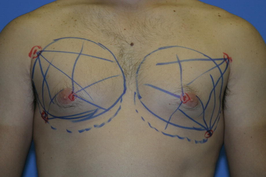
Typical preoperative markings for gynecomastia


Surgical Technique
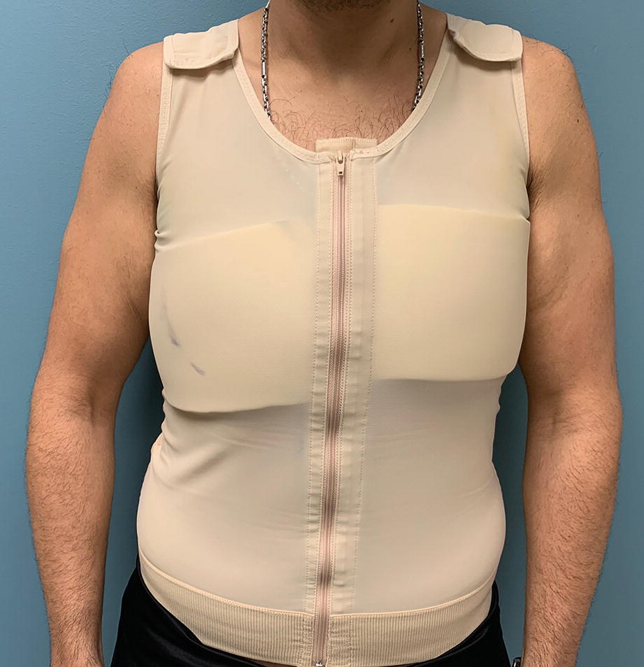

The author typically performs the procedure under general anesthesia. Access incisions extend 3–4 mm and are created with a #15 blade at the lateral inframammary fold and the inferior areolar border. The author’s wetting solution formula for general anesthesia cases is composed of 1 ml of epinephrine 1:1000 in a liter of Ringer’s lactate solution at room temperature. Infiltration of the wetting solution is performed using a power infusion pump at a rate of 300 ml per minute with even distribution throughout the breast and chest areas to be addressed. Infusion is continued to the point of tumescence including the subdermal space (typically 700–900 ml per side). Although there are “bullet ultrasonic probes” designed specifically for gynecomastia, the author finds that these probes along with the one-ring VASER probe are too aggressive and not really necessary to treat the typical gynecomastia. I employ a two-ring VASER probe (Solta Medical, Bothell, WA) at 80–90% energy level in continuous mode. The VASER exposure time is 1 minute for every 100 ml of expected total aspirate from the site (typical VASER times are 3–4 minutes per side). Aspiration is performed with 3.7 mm VentX (Solta Medical, Bothell, WA) cannulas for de-bulking and a 3 mm VentX cannula superficially. The pull-through technique for the removal of the fibro-glandular tissue is performed through the access incisions. There are a number of grasping forceps or clamps that have been described as useful for removal of gynecomastia tissue using the pull-through technique. I find tendon forceps work well in most cases and I avoid the use of sharp cannulas such as the Toledo forked cannulas because I find them too traumatic and associated with more postoperative ecchymosis. I do not hesitate to use a small curved, blunt scissor to cut some of the fibro-glandular tissue that does not readily pull through. Bleeding has not been an issue due to the significant hydrostatic pressure from the high-volume tumescence and the epinephrine effect on the tissues. Postoperative dressings include TopiFoam and a compression vest (Fig. 7.3). The surgery is performed as an outpatient procedure and patients are seen for their first visit on the third postoperative day. Depending on the volume extracted, the compression garment is worn between 1 and 2 months. Patients usually return to work after 5 days and avoid strenuous exercise for the first month.

../images/978-3-030-26875-6_7_Chapter/978-3-030-26875-6_7_Fig3_HTML.jpg

Fig. 7.3

Compression garment used postoperatively following gynecomastia surgery


Complications


During the informed consent process patients are advised of the possibility of postoperative hematoma, infection, visible scarring, nipple-areola depression deformity, contour irregularities, skin burns, and sensory changes to the nipples or breast skin. In reality complications are quite rare with this technique and the use of high amounts of epinephrine containing wetting solutions at room temperature has had a tremendous impact on avoiding the excessive bleeding and hematomas associated with the open techniques for treating gynecomastia.


Surgical Outcomes
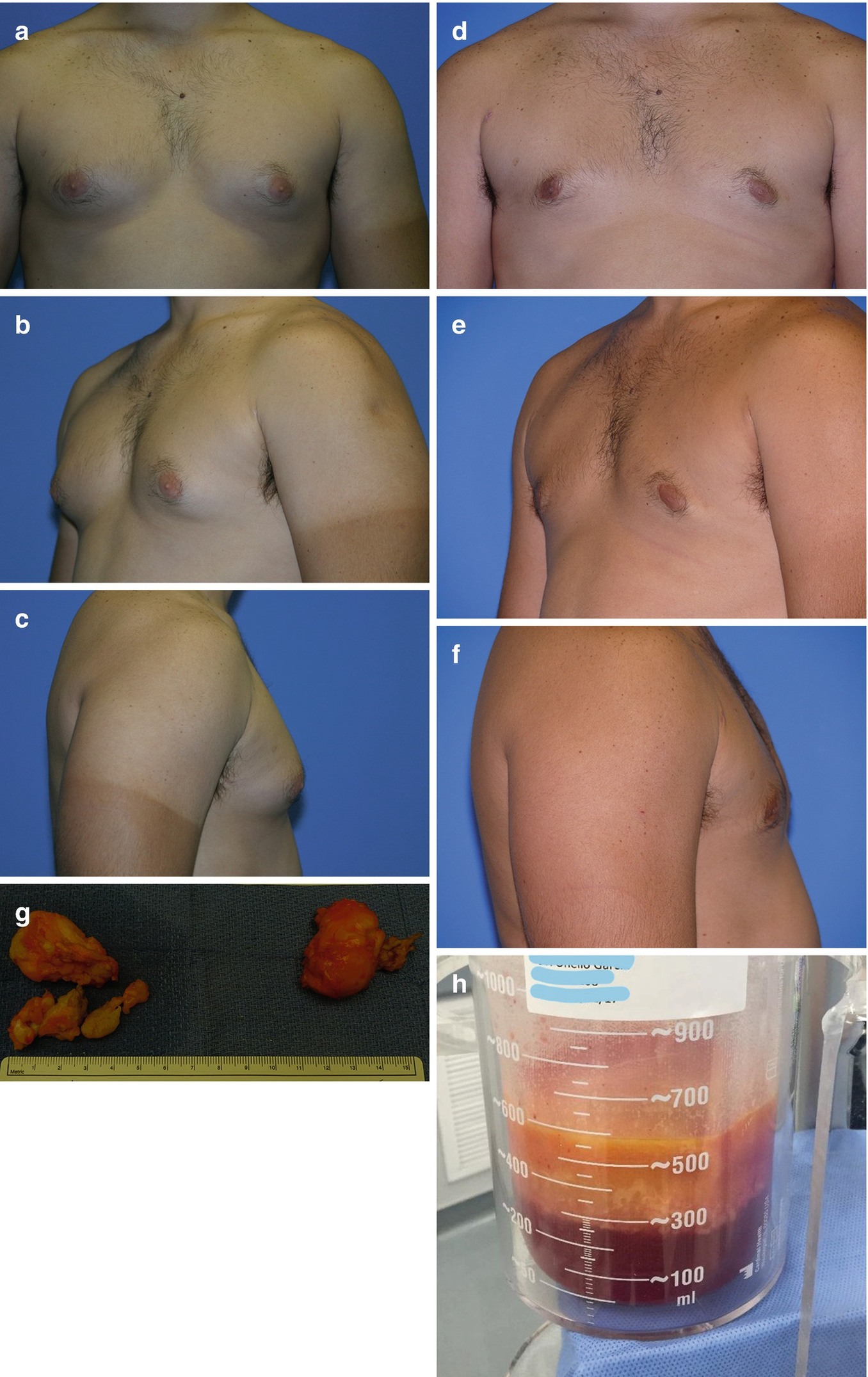

A 26-year-old male with persistent idiopathic bilateral gynecomastia was seen in consultation. VASER-assisted liposuction with resection of the subareolar fibro-glandular tissue with a pull-through technique was recommended. The surgery was performed under general anesthesia as an outpatient procedure. Wetting solution consisting of 1 ml of epinephrine 1:1000 in a liter of Ringer’s lactate solution at room temperature was infused at 300 ml per minute to a total of 750 ml per side. Ultrasound was delivered by means of a 3.7 mm, two-ring, VASER probe at 80% energy level in continuous mode for 3 minutes per breast. Aspiration was performed with a 3.7 mm VentX cannula for the deep tissue and a 3 mm VentX cannula for the superficial, subdermal liposuction. The supernatant fat aspirate volume consisted of 175 ml from each breast. Following the aspiration of the fatty tissues, the fibro-glandular component was resected via the pull-through technique. Surgical outcomes at 6 months are depicted in Fig. 7.4a–f. The subareolar glandular tissue and VASER fat aspirate are depicted in Fig. 7.4g, h.

../images/978-3-030-26875-6_7_Chapter/978-3-030-26875-6_7_Fig4_HTML.jpg

Fig. 7.4

(af) Surgical outcomes at 6 months of 26-year-old male post-VASER-assisted resection of bilateral gynecomastia. (g, h) Surgical specimen of fibro-glandular tissue and VASER aspirate
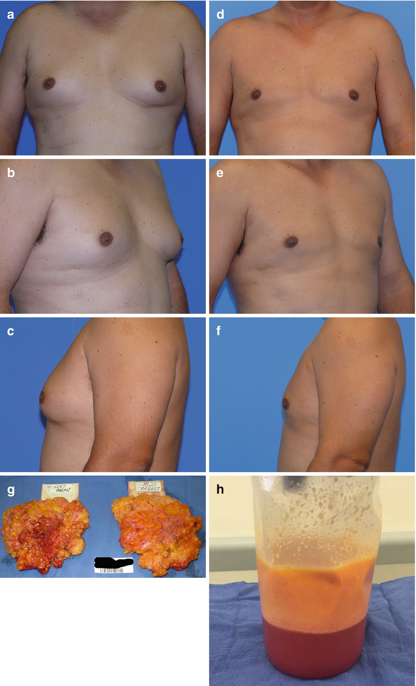

A 44-year-old, healthy, male patient with longstanding history of asymptomatic gynecomastia was seen in consultation requesting aesthetic improvement of his chest contour. VASER-assisted resection of gynecomastia was recommended. The surgery was performed under general anesthesia as an outpatient procedure. The author’s wetting solution formula for general anesthesia was infused at 300 ml per minute to a total of 800 ml per breast. A 3.7 mm, two-ring, VASER probe was employed at 90% energy level, in continuous mode for 4 minutes per breast. Aspiration was performed with a 3.7 mm VentX cannula for the deep tissue and a 3 mm VentX cannula for the superficial liposuction. Total aspirate consisted of 370 ml from right side and 300 ml from left side. The subareolar, fibro-glandular tissue was significant and could not be pulled through the 4 mm access incisions so an inferior areolar incision was used to access the tissue. Surgical outcomes at 1 year are depicted in Fig. 7.5a–f. Specimens and aspirate are depicted in Fig. 7.5g, h.

../images/978-3-030-26875-6_7_Chapter/978-3-030-26875-6_7_Fig5_HTML.jpg

Only gold members can continue reading. Log In or Register to continue

Stay updated, free articles. Join our Telegram channel

Nov 4, 2020 | Posted by in Aesthetic plastic surgery | Comments Off on Liposuction of Gynecomastia

Full access? Get Clinical Tree

Get Clinical Tree app for offline access