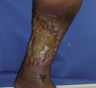
120 Leg ulcers Eri Fukaya and David J. Margolis Evidence Levels: A Double-blind study B Clinical trial ≥ 20 subjects C Clinical trial < 20 subjects D Series ≥ 5 subjects E Anecdotal case reports In general, leg ulcers are slow to heal. These wounds are on the most distal aspects of the circulatory, lymphatic, and nervous system supply and as a result there are routes for collateral vessel formation. The lower extremities are also the bearers of weight and gravity. Arterial and venous problems, infection, trauma and systemic problems such as diabetes, immunodeficiency, malnutrition or medications add to impairment. Leg ulcers secondary to other causes such as infection, basal or squamous cell cancer, or pyoderma gangrenosum can also occur but are discussed elsewhere. Management strategy Wound care All chronic wounds are contaminated but not necessarily infected. Many feel that infection can be optimally treated by debridement, which is the removal of slough and necrotic tissue to prevent and/or contain infection as well as minimizing the presence of inflammatory materials. Wound infections prolong inflammation by maintaining high levels of proinflammatory cytokines and tissue proteases. These degrade granulation tissue and delay collagen deposition and healing. Debridement and aggressive cleaning can be done mechanically with curettage, with a scissor or with a scalpel. It can also be accomplished with pressurized liquids, ultrasound, biomechanically (i.e., maggots), and enzymatically. Dressing Wound dressings should be chosen to address exudate control, wound protection, and pain relief. The frequency of dressing changes is dependent on the dressing and the reason for its use. The goal is to provide a moist wound environment to promote epithelialization. Wound exudates contain vital proteins and cytokines which facilitate autolytic debridement and promote healing. Using hydrocolloids, hydrogels, alginates, transparent films, and other bandages provide coverage and may promote the function of these enzymes. However, excessive exudate can saturate the wound bed diminishing these beneficial properties as well as macerate surrounding tissue making it more prone to injury. Finally, choosing a dressing that does not adhere to the wound bed can be important to assure that it does not disrupt re-epithelialization when it is removed. Accurately identifying the key cause of wounds is essential in the management of leg ulcers though many wounds are complex and may be of more than one etiology. Below are specific approaches to different leg ulcers. Venous leg ulcers Venous insufficiency caused by venous reflux and/or deep venous thrombosis (DVT) is a cause of ambulatory venous hypertension, which may lead to venous leg ulcers. The diagnosis for superficial or deep venous abnormality can be with a duplex ultrasound, but may also be apparent on clinical examination. If there is a high clinical suspicion for a pelvic DVT, a CT venogram should be performed. Calf muscle pump function is critical for venous outflow thus venous ulcer management hinges on good lower limb compression. The use of compression bandages for venous leg ulcer has been carefully studied and is the standard care. For prevention, it is recommended that patients wear graduated compression stockings. Leg elevation and weight reduction reduce swelling severity. Arterial ulcers Ulcers can be caused by chronic limb ischemia. Most of the affected individuals have peripheral artery disease and many have diabetes. Those with diabetes tend to have involvement of smaller arterioles. Lower limb ischemia causes pain and limb pallor. Arterial circulation can be evaluated non-invasively with ankle brachial index (ABI) and pulse volume recordings (PVR). The PVR may be non-diagnostic in patients with advanced calcification such as those with diabetes. In this setting a duplex ultrasound or CT angiography is necessary. Treatment including proximal obstructive lesions causing ischemia may require invasive interventions such as angioplasty or arterial bypass grafting. Patients with poor arterial circulation can develop deep soft tissue infection, osteomyelitis, or gangrene leading to amputation, thus close monitoring is essential. Only gold members can continue reading. Log In or Register to continue Share this: Click to share on X (Opens in new window) X Click to share on Facebook (Opens in new window) Facebook Related Related posts: Mucoceles Tinea capitis Herpes genitalis Necrolytic migratory erythema Nevoid basal cell carcinoma syndrome Rocky Mountain spotted fever and other rickettsial infections Stay updated, free articles. Join our Telegram channel Join Tags: Treatment of Skin Disease Comprehensive Therapeutic Strategies Aug 7, 2016 | Posted by admin in Dermatology | Comments Off on Leg ulcers Full access? Get Clinical Tree
120 Leg ulcers Eri Fukaya and David J. Margolis Evidence Levels: A Double-blind study B Clinical trial ≥ 20 subjects C Clinical trial < 20 subjects D Series ≥ 5 subjects E Anecdotal case reports In general, leg ulcers are slow to heal. These wounds are on the most distal aspects of the circulatory, lymphatic, and nervous system supply and as a result there are routes for collateral vessel formation. The lower extremities are also the bearers of weight and gravity. Arterial and venous problems, infection, trauma and systemic problems such as diabetes, immunodeficiency, malnutrition or medications add to impairment. Leg ulcers secondary to other causes such as infection, basal or squamous cell cancer, or pyoderma gangrenosum can also occur but are discussed elsewhere. Management strategy Wound care All chronic wounds are contaminated but not necessarily infected. Many feel that infection can be optimally treated by debridement, which is the removal of slough and necrotic tissue to prevent and/or contain infection as well as minimizing the presence of inflammatory materials. Wound infections prolong inflammation by maintaining high levels of proinflammatory cytokines and tissue proteases. These degrade granulation tissue and delay collagen deposition and healing. Debridement and aggressive cleaning can be done mechanically with curettage, with a scissor or with a scalpel. It can also be accomplished with pressurized liquids, ultrasound, biomechanically (i.e., maggots), and enzymatically. Dressing Wound dressings should be chosen to address exudate control, wound protection, and pain relief. The frequency of dressing changes is dependent on the dressing and the reason for its use. The goal is to provide a moist wound environment to promote epithelialization. Wound exudates contain vital proteins and cytokines which facilitate autolytic debridement and promote healing. Using hydrocolloids, hydrogels, alginates, transparent films, and other bandages provide coverage and may promote the function of these enzymes. However, excessive exudate can saturate the wound bed diminishing these beneficial properties as well as macerate surrounding tissue making it more prone to injury. Finally, choosing a dressing that does not adhere to the wound bed can be important to assure that it does not disrupt re-epithelialization when it is removed. Accurately identifying the key cause of wounds is essential in the management of leg ulcers though many wounds are complex and may be of more than one etiology. Below are specific approaches to different leg ulcers. Venous leg ulcers Venous insufficiency caused by venous reflux and/or deep venous thrombosis (DVT) is a cause of ambulatory venous hypertension, which may lead to venous leg ulcers. The diagnosis for superficial or deep venous abnormality can be with a duplex ultrasound, but may also be apparent on clinical examination. If there is a high clinical suspicion for a pelvic DVT, a CT venogram should be performed. Calf muscle pump function is critical for venous outflow thus venous ulcer management hinges on good lower limb compression. The use of compression bandages for venous leg ulcer has been carefully studied and is the standard care. For prevention, it is recommended that patients wear graduated compression stockings. Leg elevation and weight reduction reduce swelling severity. Arterial ulcers Ulcers can be caused by chronic limb ischemia. Most of the affected individuals have peripheral artery disease and many have diabetes. Those with diabetes tend to have involvement of smaller arterioles. Lower limb ischemia causes pain and limb pallor. Arterial circulation can be evaluated non-invasively with ankle brachial index (ABI) and pulse volume recordings (PVR). The PVR may be non-diagnostic in patients with advanced calcification such as those with diabetes. In this setting a duplex ultrasound or CT angiography is necessary. Treatment including proximal obstructive lesions causing ischemia may require invasive interventions such as angioplasty or arterial bypass grafting. Patients with poor arterial circulation can develop deep soft tissue infection, osteomyelitis, or gangrene leading to amputation, thus close monitoring is essential. Only gold members can continue reading. Log In or Register to continue Share this: Click to share on X (Opens in new window) X Click to share on Facebook (Opens in new window) Facebook Related Related posts: Mucoceles Tinea capitis Herpes genitalis Necrolytic migratory erythema Nevoid basal cell carcinoma syndrome Rocky Mountain spotted fever and other rickettsial infections Stay updated, free articles. Join our Telegram channel Join Tags: Treatment of Skin Disease Comprehensive Therapeutic Strategies Aug 7, 2016 | Posted by admin in Dermatology | Comments Off on Leg ulcers Full access? Get Clinical Tree