Lasers, injectable fillers, and neurotoxins are widely used in facial restoration and rejuvenation by a variety of practitioners. Although they are less invasive than traditional surgical modalities, they still carry risks for both transient as well as permanent complications. It is paramount for the practitioner to understand these complications, optimize their prevention, and initiate appropriate treatment when they are encountered. This article reviews early, often transient, complications as well as delayed, often prolonged or permanent, complications, with particular focus on prevention and management.
Key points
- •
Lasers, injectable fillers, and neurotoxins are extensively used in facial aesthetics.
- •
Fractional laser resurfacing has introduced a new era in skin rejuvenation, with a lower complication rate and shorter downtime.
- •
The applications for fillers and neurotoxins have expanded beyond the traditional nasolabial fold correction and forehead lines.
- •
Permanent and problematic complications can occur, and prompt recognition and appropriate management minimize the sequelae.
- •
It is the practitioner’s understanding of facial anatomy and aesthetic principles that translates the use of these modalities into a safe and pleasing result.
Introduction
Lasers, injectable fillers, and neurotoxins are widely used in facial restoration and rejuvenation by a variety of practitioners. Although they are less invasive than traditional surgical modalities, they still carry risks for both transient as well as permanent complications. It is paramount for the practitioner to understand these complications, optimize their prevention, and initiate appropriate treatment when they are encountered. This article reviews both early, often transient, complications as well as delayed, often prolonged or permanent, complications with particular focus on prevention and management.
Laser technologies: sequelae and complications
Applications for laser technologies in the practice of facial plastic surgery have expanded. There are several effective modalities for the treatment of aging skin, vascular lesions, telangiectasia, pigmentary irregularities, and acne scarring. Lasers deliver light energy that is absorbed by target tissues (chromophores) to produce photothermal injury. The penetration and energy of the laser, as well as the absorption and thermal relaxation characteristics of the chromophores, dictate the induced thermal injury. In high-energy ablative lasers, the thermolysis involves both the epidermis and the dermis, whereas in low-energy nonablative lasers an intact epidermis cover is spared. The introduction of fractionation has improved the safety of laser resurfacing by using micro pinpoint ablations and allowing the healthy surrounding tissue to accelerate healing. Although laser complications can occur following any treatment, there are specific laser and patient factors that contribute to their occurrence ( Table 1 ). The American National Standard Institute provides comprehensive details on the safe use of lasers in health care, and these recommendations should be consulted when setting up a laser procedure room. Standard laser precautions include:
- •
Adequate signs
- •
Protective eyewear
- •
Routine device check with pretesting
| Complication | Risk Factors | Prevention and Management |
|---|---|---|
| Erythema | Sensitive and thin skin Excessive sun damage | Icing, rigorous sun precautions Masking with makeup Treat with topical Biafine (Valeant, Montreal, QC) and 590-nm LED photomodulation Consider a mild topical corticosteroid if condition persists |
| Blistering and burns | High-energy/penetration lasers Improper pulse stacking or high-density passes Insufficient cooling of the dermis Loss of pain feedback with heavy sedation or general anesthesia | Implement standard device safety and review of laser settings Allow the dermis to cool between passes and after treatment Burn care for severe thermal injuries |
| Infection and herpetic eruption | Closed facial dressing left >48 h Insufficient facial hygiene/care History of herpetic rash | Adherent to posttreatment facial care with topical disinfectant with hypochlorous acid 0.01% (NeutroPhase, NovaBay, Emeryville, CA) or acetic acid 0.25%–0.0125% (vinegar solution) Prophylactic antibiotic (cephalosporin), and a 1–2 wk anti viral course (valacyclovir) started 24–48 h before treatment |
| Acne and milia | Closed facial dressings Oil-based creams | Daily facial rinses and noncomedogenic moisturizer Course of oral tetracycline for persistent acne Milia can benefit from topical tretinoin, gentle epidermabrasion, or extraction |
| Postinflammatory hyperpigmentation | Fitzpatrick skin type III–VI Recent sun exposure/tanning History of hyperpigmented healing | Careful skin type selection with appropriate laser type setting Sun precaution 2–4 wk before treatment and continued for 2–4 mo Prophylactic or therapeutic 2%–4% hydroquinone, 2%–4% kojic acid or Kligman formula (5% hydroquinone, 0.1% tretinoin, and 0.1% dexamethasone). This should be started 2–4 wk before treatment and continued for 2–4 mo |
| Scarring and hypertrophic healing | Secondary to infections or burns Poor healing capacity History of keloid formation The periorbital region, the neck, and off-face areas | Scar-prone areas require lower fluence and density Apply silicon gel dressing to healing scars and hypertrophic bands Intralesion corticosteroid or 5-fluorouracil |
There is a risk for ignition whenever high-energy lasers are in use. Accordingly:
- •
Patients should not have flammable hair sprays or cosmetic products.
- •
Oxygen supplementation should not flow close to the laser field.
- •
All flammable tubing should be protected with moist towels.
- •
Saline should be readily available.
Bruising, Erythema, and Edema
Acute inflammatory skin reaction is an expected sequela following laser treatment. This reaction is generally short-lived and resolves within 2 to 4 days following nonablative treatments and 2 to 4 weeks following ablative treatments. Prolonged erythema beyond these periods is seen in about 1% and 10% of patients, respectively. Sensitive plethoric skin, rosacea, and light skin color are more prone to redness. Although women can mask erythema easily with makeup, this is not an attractive option for men. Topical Biafine (Valeant, Montreal, QC) and light-emitting diode photomodulation seem to improve persistent symptoms. Hypersensitivity to topical or systemic treatments, albeit difficult to diagnose, should be suspected whenever pruritus or other allergic symptoms are present. Focal edema and exudates can result, leading to crust formation with small skin breaks, which delays reepithelialization. This problem can be minimized with early icing and liberal moisturizing. Although bruising is rare following routine laser treatment, pulse dye laser is the exception, and patients should be forewarned to better plan the timing of their treatment.
Blistering and Burns
Small blistering occurs particularly with the use of yttrium aluminum garnet (YAG) and Q-switched lasers. These blisters are typically 1 to 2 mm in diameter and can cover the treated area. However, this invariably resolves with no sequelae in 2 to 4 days. Large blisters are more concerning and can indicate significant thermal injury. Insufficient cooling or failure of the cooling system can result in burns of various severities ( Fig. 1 ), typically as parallel patches taking the shape of the headpiece. Severe pain is an early indication that heating is excessive, but this important feedback is lost if the procedure is performed under deep sedation or a general anesthetic. Prompt icing and good skin care with daily antiseptic washes can improve the healing of blisters and superficial burns. More extensive injuries are managed in a similar fashion to general burn care.
Infection and Herpetic Eruption
The most common infection after laser skin resurfacing is the reactivation of herpes simplex virus (HSV), with reported rates ranging from 0.5% to 5%. The presence of active lesions is a contraindication to laser treatment. A patient’s history of prior HSV eruptions seems unreliable for risk identification. Primary bacterial infections can also occur during the reepithelialization phase. Staphylococcus aureus followed by Pseudomonas aeruginosa are the most common culprits. Candida fungal infections are rare, but have been reported. Many practitioners initiate prophylactic antiviral treatment regardless of the HSV history status. A 1-week to 2-week course of valacyclovir, started 24 to 48 hours before treatment, can decrease the risk for herpetic eruption to less than 0.5%. Some practitioners also initiate prophylactic oral antibiotics (cephalosporin) for 1 week. Spreading cellulitis, purulent discharge, or constitutional symptoms should prompt the physician to obtain cultures for guided antimicrobial therapy. Good facial hygiene is always a critical component of postresurfacing treatment. Open dressing is preferred, because it has the advantage of facilitating early facial care. If a closed dressing is used with deeper resurfacing, it should not be left for more than 48 hours because this increases bacterial growth. The patient should use antiseptic rinses with hypochlorous acid 0.01% (NeutroPhase, NovaBay, Emeryville, CA) or acetic acid 0.25% to 0.0125% (vinegar solution) several times a day until reepithelialization is complete.
Acne and Milia
Acneiform rash erupts following fractional laser treatment in 5% to 10% of cases. Milia represent entrapped hair follicles or sebaceous glands under the reepithelializing skin and usually appear 2 to 3 weeks after treatment. Both conditions seem to be exacerbated with closed facial dressings and oil-based creams. Starting daily facial rinses and noncomedogenic moisturizer can improve these conditions. For patients with a persistent acne flare, a course of oral tetracycline is suitable. Milia can benefit from topical tretinoin, gentle epidermabrasion, or extraction.
Dyschromia
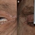
Postinflammatory hyperpigmentation is probably the most common delayed complication ( Fig. 2 ) and is rarely permanent. It is typically encountered several weeks following ablative CO 2 or Er:YAG laser resurfacing, because the inflammatory healing response leads to epidermal or dermal hypermelanosis. Although all skin types can show this inflammatory response, it is almost expected in Fitzpatrick skin types III and greater. The healing behavior of previous wounds or acne sites can also assist in predicting the individual’s tendency for hyperpigmented healing. All patients should have strict sun precautions at least 2 to 4 weeks before treatment and continued for at least 2 to 4 months. In high-risk patients, a topical bleaching regimen such as 4% hydroquinone, 2% kojic acid, or Kligman formula (5% hydroquinone, 0.1% tretinoin, and 0.1% dexamethasone) can markedly quiesce melanocytes and is useful for both prevention as well as treatment. Once the desired effect is achieved, a gradual tapering off can minimize rebound. Although hyperpigmentation is temporary, it can persist for several months despite topical treatment. Such patients often show remarkable improvement with superficial chemical peels.
Hypopigmentation is a rare delayed complication that can become evident even several months following treatment and may become permanent. Darker skin tone, previous history of resurfacing, or a prior deep peel are well-recognized risk factors. Demarcation lines, indicating hypopigmentation of the treated skin, were frequently seen with full ablative resurfacing (see Fig. 1 ). However, this is rare in the fractionation era because of the sparing of healthy skin between ablation zones. It is best to address all the aesthetic units of the face with feathering at the borders. This approach provides a smooth transition without abrupt demarcation lines or evident differences in skin texture. Spontaneous repigmentation often takes place over 3 to 4 months. In cases with permanent hypopigmentation, excimer laser, topical photochemotherapy, or alternatively micropigment makeup are all suitable options.
Textural Irregularities
The treatment of isolated facial aesthetic units can accentuate differences in skin texture; this is often seen as smoother, tighter skin in an aggressively treated midface region contrasting with more relaxed and irregular skin over periorbital or lower face regions (see Fig. 1 ). Evident depressions can develop in areas with thin skin and subcutaneous tissue if the deep thermal injury prevents fibroblasts from generating and remodeling collagen. Topical corticosteroids could be a risk factor by suppressing inflammation and fibroblast activity. Infrared or visible-light laser can improve soft irregularities persisting after 6 months by promoting nonablative dermal remodeling. In contrast, autologous fat transfer, dermal fillers, and subcision are better options for prominent depressions.
Scarring and Hypertrophic Healing
This is a rare complication that is caused by excessive thermal injury or poor healing capacity, or it may be secondary to an infectious complication (see Fig. 1 ). Important patient risk factors include keloid formation, smoking, chronic conditions, or medications that interfere with wound healing. Overly aggressive fluences, high density, insufficient cooling between passes, or improper pulse stacking are all technical factors that can lead to scarring. The periorbital region, the neck, and off-face areas are sensitive and require lower fluence and density. If a hypertrophic or keloid scar develops, silicon gel dressing, intralesion corticosteroid, or 5-fluorouracil injection can improve the cosmetic result.
Injectable fillers: sequelae and complications
A wide variety of injectable materials serve to restore facial volume and soften static wrinkles or folds. It is important for the practitioner to be familiar with the characteristics of each filler and its appropriate use and precautions ( Table 2 ). The aesthetic goal for each patient is an individual preference, and should be discussed carefully before treatment. Although many patients come with well-formed desires, it is the practitioner’s understanding of facial aging and aesthetic principles that translates the use of fillers into a rejuvenated and pleasing result. Digital photography can aid in documenting pretreatment status, highlighting asymmetries, and detailing facial analysis and goals. In general, complications can arise either from injection trauma or from the filler material. Hyaluronic acid (HA) is the only filler that can be quickly and safely dissolved with enzymatic injection if there is a complication or an undesired result. Hyaluronidase has been used off-label for the last several years with excellent results. Two products approved by the US Food and Drug Administration (FDA) have recently entered the market: Vitrase (Bausch & Lomb, Rochester, NY) and Hylenex (Halozyme Therapeutics, San Diego, CA). This advantage of HA fillers remains significant, particularly for practitioners who are still developing their techniques.
Related posts:

Stay updated, free articles. Join our Telegram channel

Full access? Get Clinical Tree


