Laparoscopic Surgery for Stress Urinary Incontinence and Pelvic Organ Prolapse

17 Laparoscopic Surgery for Stress Urinary Incontinence and Pelvic Organ Prolapse


Since laparoscopic retropubic urethropexy was introduced in 1991, laparoscopic access and techniques have been applied to most abdominal-route and numerous vaginal-route surgical procedures for treatment of urinary incontinence and pelvic organ prolapse. Adoption of laparoscopic sacral colpopexy has increased in the past decade and has recently evolved to robotic assistance. The possible advantages of laparoscopic surgery are improved visualization of anatomy of the space of Retzius and peritoneal cavity because of laparoscopic magnification, insufflation effects, and improved hemostasis; shortened hospitalization resulting in potential cost reduction; decreased postoperative pain and more rapid recovery and return to work; and better cosmetic appearance of smaller incisions. Disadvantages of laparoscopic surgery include a steep learning curve in acquiring suturing skills, technical difficulty of retroperitoneal dissection, increased operating time early in the surgeon’s experience, and possible greater hospital cost secondary to increased operating room time and the use of disposable surgical instruments. These listed disadvantages, inadequate experience in advanced laparoscopy in residency and fellowship programs, and recently introduced minimally invasive midurethral sling and apical suspension procedures have thwarted widespread adoption of laparoscopic surgery for urinary incontinence and prolapse. The literature regarding these procedures consists of many case series from surgeons subspecializing in advanced laparoscopy. Comparative, adequately powered studies regarding laparoscopic surgery for stress urinary incontinence and prolapse are rare.


LAPAROSCOPIC RETROPUBIC SURGICAL PROCEDURES


In the first report, Vancaillie and Schuessler (1991) laparoscopically duplicated the conventional Marshall-Marchetti-Krantz (MMK) procedure. Subsequently, Albala et al. (1992) published a case series of MMK and Burch procedures. Many investigators have modified the laparoscopic retropubic colposuspension using varying numbers and types of suture, synthetic mesh, staples, bone anchors, coils, tacks, fibrin sealant, and radiofrequency. Various suturing and needle devices have been used to simplify laparoscopic suturing and knot-tying, the most difficult skills to acquire.


The Burch procedure can be performed via a small laparotomy incision with good long-term success and minimal morbidity. To replace this accepted and effective approach with laparoscopic access, there must be comparable efficacy and an equivalent or better complication rate. If one is to consider performing a laparoscopic retropubic bladder neck suspension, we believe that it should be performed exactly the same as the open procedure. Modifications to the open procedure represent essentially new operations, thus requiring further study and outcome analysis.


Anatomy


Thorough knowledge of the anatomy of the anterior abdominal wall is mandatory for safe and effective trocar insertion. The umbilicus is approximately at the L3–L4 level, and the aortic bifurcation is at L4–L5. In obese women the umbilicus is caudal to the bifurcation. Thus, the intraumbilical trocar should be introduced at a more acute angle toward the pelvis in thin women and closer to 90 degrees in obese women. The left common iliac vein courses over the lower lumbar vertebras from the right side and may be inferior to the umbilicus. Common iliac arteries course 5 cm before bifurcating into the internal and external iliac arteries. The ureter crosses the common iliac artery at or above its bifurcation.


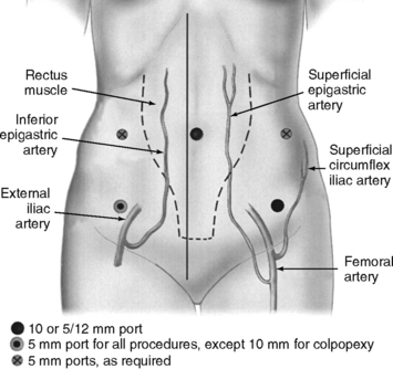
The superficial epigastric artery, a branch of the femoral artery, courses cephalad and can be transilluminated. The inferior epigastric artery branches from the external iliac artery at the medial border of the inguinal ligament and runs laterally to and below the rectus sheath at the level of the arcuate line. Two inferior epigastric veins accompany this artery (Fig. 17-1). The median umbilical ligament, the embryonic urachus, is attached to the apex of the bladder and extends to the umbilicus. The urachus remains patent in some women and may be somewhat vascular. The medial umbilical folds, the peritoneum overlying the obliterated umbilical arteries, are the lateral landmarks of dissection of the parietal peritoneum during transperitoneal surgery into the space of Retzius. The upper margin of the dome of the bladder is noted approximately 3 cm above the pubic symphysis when the bladder is filled with 300 mL of fluid. Before distention of the bladder, the upper margin of the dome lies several centimeters above the pubic symphysis.


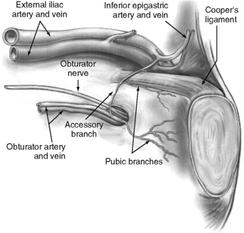
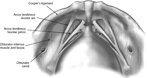
The important landmarks of the space of Retzius are Cooper’s ligaments; the accessory or aberrant obturator veins; the obturator neurovascular bundles, which are 3 to 4 cm above the arcus tendineus fasciae pelvis; the bladder neck, which is delineated by placing traction on the Foley bulb; and the arcus tendineus fasciae pelvis and arcus tendineus levator ani, which insert into the pubic bone (Figs. 17-2 and 17-3).


Operative Technique


OPERATIVE SETUP AND INSTRUMENTATION


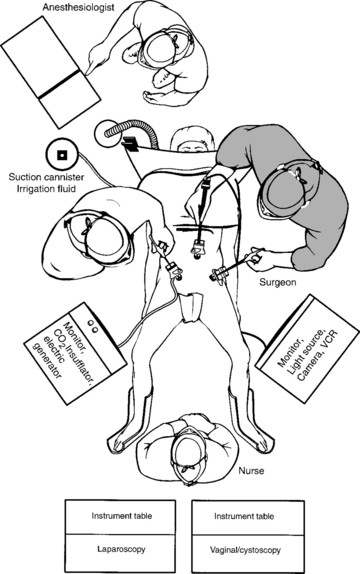
The operating room setup is shown in Figure 17-4. The monitor screens should be placed laterally to the legs in direct view of the surgeon standing on the opposite side of the table. The scrub nurse should be in the center if two monitor screens are used; otherwise, the scrub nurse is located behind one surgeon and the electrosurgical unit or harmonic scalpel on the opposite side. After the three-way Foley catheter and uterine manipulator (if needed) have been placed, the vaginal tray with cystoscope is set aside, if desired, for later use.


Ideal stirrups for combined laparovaginal cases are the Allen stirrups and Yellofins (Allen Medical Systems, Acton, MA) that have levers that can quickly convert the patient from low to high lithotomy position while preserving sterility of the field. A sterile pouch attached to each thigh is equipped with commonly used instruments, such as unipolar scissors, bipolar cautery, graspers, and laparoscopic blunt-tipped dissectors. The irrigation should be set up before making incisions for trocars.


For standard suturing technique, needle holder preference is determined by comfort of the surgeon. Conventional and 90-degree self-righting German needle holders (Ethicon Endo-Surgery, Inc., Cincinnati, OH) have ratchet spring handles, and the Talon curved needle drivers with spring handles (Cook OB/GYN, Spencer, IN) self-right the needle at an angle, either 45 or 90 degrees to the needle driver shaft, depending on the style chosen. The Storz Scarfi needle holder and notched assistant needle holder (Karl Storz Endoscopy, Culver City, CA) are most like conventional needle holders used during laparotomy. However, the handles are difficult to maintain and may pop open after extended use. The needle holder tips may become magnetized, which hampers needle grasping. Disposable suturing devices have been introduced that include the Endo-stitch (U.S. Surgical Corp., Norwalk, CT) and the Capio CL (Cooper’s ligaments) (Microvasive Boston Scientific, Inc., Natick, MA). Extracorporeal knot-tying is preferred because of technical facility and the ability to hold more tension on the suture. The choice of an open-ended or close-ended knot pusher for extracorporeal knot-tying depends on surgeon preference. Our suture of choice is the double-armed No. 0 Ethibond 36-inch suture on an SH or CT-2 needle (Ethicon, Inc., Somerville, NJ). Our alternative choice for suture is No. 0 Gore-Tex (W.L. Gore & Associates, Inc., Phoenix, AZ). A 48-inch suture is preferred when suturing from ports at the level of the umbilicus. Sterile steel thimbles may be used by the surgeon or assistant when elevating the vagina while the surgeon is placing the stitches in the vaginal wall.


SKIN INCISIONS FOR TROCAR SITES


Intraumbilical or infraumbilical incisions are made depending on the anatomy of the umbilicus. Many variations of the accessory trocar sites have been described. We use two additional trocars: a 5/12-mm disposable trocar with reducer in the right lower quadrant (if knot-tying from the right) lateral to the right inferior epigastric vessels and a reusable 5-mm port or an additional 5/12-mm disposable trocar, with reducer in the left lower quadrant lateral to the left inferior epigastric vessels. Trocars are placed laterally to the rectus muscle, approximately 3 cm medial to and above the anterior superior iliac spine. Based on an anatomic study by Whiteside et al. (2003), we know that ilioinguinal and iliohypogastric nerve entrapment during fascial closure may be reduced if the ports are placed at least 2 cm cephalad to the anterior superior iliac spines. An additional 5-mm port may be placed on the principal surgeon’s side so that he or she can operate with two hands. Both reusable and disposable ports may be secured with circumferential screws to prevent port slippage. Versa Step Plus trocars (U.S. Surgical Corp., Norwalk, CT) allow easy introduction of needles, maintain pneumoperitoneum during extracorporeal knot-tying, and prevent port slippage because of the expandable sleeve. Port placement is shown in Figure 17-1.


ROUTE: EXTRAPERITONEAL OR INTRAPERITONEAL


The choice of extraperitoneal or intraperitoneal approach depends on whether concomitant intraperitoneal procedures are being performed, on whether the patient has had previous abdominal wall surgery, and on surgeon preference. Previous retropubic surgery is a contraindication for extraperitoneal approach, and low transverse or midline incisions make the dissection more difficult and prone to failure. Some surgeons report less operating time, easier dissection, and fewer bladder injuries with the extraperitoneal route. This route is sometimes easier because the balloon performs the majority of the dissection. We prefer the intraperitoneal approach because it allows a larger operating space for safe, secure, comfor table handling of the suture. Furthermore, a culdoplasty or other intraperitoneal surgery can be performed concomitantly.


The intraperitoneal approach begins with insertion of the 0-degree laparoscope (5 mm or 10 mm) through a respective 5- or 10-mm intraumbilical or infraumbilical cannula followed by intra-abdominal insufflation. Inspection of the peritoneal cavity is performed, delineating the inferior epigastric vessels, abdominal and pelvic organs, pelvic adhesions, and coexisting abdominal or pelvic pathology. Two additional trocars (a 5-mm and a 5/12-mm or two 5/12-mm ports) are placed under direct vision, one on each side, as previously noted.


All trocars are nondisposable except the 5/12-mm trocar through which 5- and 10-mm instruments are introduced. This site is used for introduction of the needles and sutures. Some surgeons backload the suture through 5-mm ports and introduce and remove needles through the skin incisions. This is easily accomplished in thinner patients. However, trauma to the subcutaneous tissues and inferior epigastric vessels may result with this technique. Furthermore, it is difficult to use this technique with sutures with double-armed needles. The bladder is filled with 200 to 300 mL sterile water or saline (indigo carmine or methylene blue is optional). Using sharp dissection with electrocautery or harmonic scalpel, a transverse incision 2 cm above the bladder reflection between the medial umbilical folds is made. Identification of the loose areolar tissue at the point of incision confirms a proper plane of dissection. Blunt and sharp dissection aiming toward the posterior-superior aspect of the pubic symphysis decreases risk of bladder injury. Blunt dissection is then carried out inferolaterally on both sides to identify the pubic symphysis, Cooper’s ligaments, and bladder neck. Medial dissection over the urethra should be avoided.


The extraperitoneal approach to the space of Retzius is best performed using a balloon dissector (Origin Medsystems, Menlo Park, CA; U.S. Surgical Corp., Norwalk, CT). This approach begins with an infraumbilical incision and modified open laparoscopy. After the anterior sheath of the rectus fascia is incised, a finger is swept around the rectus muscle over the posterior rectus sheath and into the preperitoneal space. Lubricated Hagar dilators can aid blunt dissection to the retropubic space. The space of Retzius is dissected by tunneling the tip of the dissector to the posterior superior aspect of the pubic symphysis. The balloon is subsequently inflated under video guidance. A 10-mm Hasson cannula or its modification (some alternatives have infla table balloons on the shaft to decrease CO2 loss) is then placed, a 0-degree laparoscope is inserted, and CO2 is insufflated into the preperitoneal space.


Further delineation of the retropubic anatomy is achieved with blunt dissection. Two additional ports are placed under direct vision laterally to the inferior epigastric vessels, taking special care to avoid entry into the peritoneal cavity.


LAPAROSCOPIC BURCH COLPOSUSPENSION


After the space of Retzius is exposed, the surgeon places two fingers in the vagina and identifies the urethrovesical junction by placing gentle traction on the Foley catheter. With elevation of the vaginal fingers, the vaginal wall lateral to the bladder neck is exposed by using a laparoscopic blunt-tipped dissector. As recommended by Tanagho (1976), no dissection is performed within 2 cm of the bladder neck to avoid bleeding and damage to the periurethral musculature and nerve supply.


We place stitches in the vaginal wall, excluding the vaginal epithelium at the level of, or just proximal to, the midurethra and bladder neck (see Fig. 17-3). No. 0 nonabsorbable suture is placed in a figure-of-eight stitch incorporating the entire thickness of the anterior vaginal wall. The needle is then passed ipsilaterally through Cooper’s ligaments. If double-armed suture is used, we make two passes through Cooper’s ligaments and subsequently tie above the ligaments. We place Gelfoam (Pharmacia Upjohn, Inc., Kalamazoo, MI) between the vaginal wall and the obturator fascia before knot-tying to promote fibrosis. With simultaneous vaginal elevation, the suture is tied with six extracorporeal square knots. Two granny half-hitches (equivalent to a surgical knot) and a flat square knot secure the stitch. Our technique for laparoscopic Burch procedure is illustrated in Color Plate 3.


Sutures are tied as they are placed to avoid tangling. Midurethral stitches are placed first, although this is a matter of preference. Placing stitches from the contralateral port is easier. For example, a right-handed surgeon elevates the vagina with his or her left hand while simultaneously placing stitches on the patient’s right side through the lower left port. In this circumstance, the principal surgeon must switch sides with the assistant. If the lower quadrant ports are placed higher (at or slightly below the level of the umbilicus), placement of ipsilateral stitches is facilitated because the angle to the ipsilateral vaginal wall and Cooper’s ligaments is less acute. The appropriate level of bladder neck elevation is estimated with the assistant’s vaginal hand. The assistant elevates the vaginal wall to place the urethra and bladder neck in a high retropubic position, which does not result in kinking or compression of the urethra. The goal is to elevate bilaterally the vaginal wall to the level of the arcus tendineus fasciae pelvis so that the bladder neck is supported and stabilized by the vaginal wall that acts as a hammock between both “white lines.” In tying the sutures, the surgeon should not reapproximate the vaginal wall to Cooper’s ligaments or place too much tension on the vaginal wall. A suture bridge of 1.5 to 2 cm is common.


When double-armed needles are used, application of two 5/12-mm ports can streamline the procedure. A surgeon can use one port for suture and needle introduction and the other port for needle exit and knot-tying. After introducing the first needle, the surgeon takes a stitch through the endopelvic fascia and vaginal wall, then through Cooper’s ligaments, and removes it through the opposite port. The second needle is introduced and an additional stitch is taken perpendicularly in the endopelvic fascia to the first stitch (resulting in a double throw), then through Cooper’s ligaments. The second stitch is removed and extracorporeal knot-tying is performed after placement of Gelfoam. This technique diminishes suture locking. Gore-tex suture is single armed and tends to slip through the tissue much easier than braided polyester suture.


LAPAROSCOPIC PARAVAGINAL DEFECT REPAIR


The laparoscopic approach to the space of Retzius for the paravaginal defect repair is identical to the Burch procedure. The dissection is carried out laterally with a blunt-tipped dissector until the obturator internus muscle, obturator foramen with neurovascular bundle, and arcus tendineus fasciae pelvis are delineated. A vaginal hand is used to elevate the vagina and medially retract the bladder, thus aiding in the dissection of the vagina and lateral landmarks. Blunt dissection is carried out dorsally until vaginal palpation of the ischial spine is visualized laparoscopically.


Starting at the vaginal apex, a No. 2–0 nonabsorbable, 36- or 48-inch suture on a CT-2 needle is used to place a stitch into the full thickness of the vagina (excluding the vaginal epithelium) and then into the arcus tendineus fasciae pelvis, which is 3 to 4 cm below the obturator fossa (Color Plate 4). This is then tied extracorporeally. An additional three to five sutures are placed through the vaginal wall and into the arcus tendineus or fascia of the obturator internus muscle at 1- to 2-cm intervals until the defect is closed. The same procedure is performed on the opposite side.


If this procedure is performed concomitantly with the Burch colposuspension, the paravaginal defect repair should be performed first because exposure of the lateral defect decreases after the Burch sutures are tied. We first place the stitch at the level of the ischial spine and then place subsequent distal stitches as needed.


GENERAL INTRAOPERATIVE AND POSTOPERATIVE PROCEDURES


The patient is instructed to take one bottle of magnesium citrate or equivalent bowel preparation and limit her diet to clear liquids on the day before surgery. Placing an orogastric or nasogastric tube to decompress the stomach at the time of surgery is also helpful. Patients receive prophylactic, intravenous antibiotic therapy 30 minutes before surgery. Pneumatic compression stockings are routinely used. The Burch procedure and paravaginal defect repair are performed under general anesthesia in the low lithotomy position. A 16-French three-way Foley catheter with a 20- to 30-mL balloon is attached to continuous drainage, and the irrigation port is connected to sterile water or saline.


After all sutures are placed and tied, transurethral cystoscopy or suprapubic telescopy is done to document ureteral patency and absence of sutures in the bladder. A suprapubic catheter is placed, if desired. The surgeon must reinspect the space of Retzius for bleeding while reducing the carbon dioxide insufflation. Routine closure of the peritoneum is performed based on surgeon preference. All ports are removed under direct visualization, and the peritoneum and fascia of all 10- or 12-mm incisions are reapproximated with the fascial closure instrument (Karl Storz, Tuttingen, Germany) or the Grice needle (New Ideas in Medicine, Inc., Clearwater, FL). The skin is closed in a subcuticular fashion. The fascia and subcutaneous fat are infiltrated with a long-acting local anesthetic, such as bupivacaine hydrochloride 0.5%, if desired.


Postoperative care consists of oral pain medication (intravenous, if needed), rapid diet advancement, ambulation, and continuation of pelvic muscle exercises. Voiding trials begin as soon as the patient is ambulatory. Intermittent self-catheterization protocols can begin immediately, especially if the patient was preoperatively taught the technique. Alternatively, the suprapubic tube is clamped when the patient is awake. The patient voids with urge or every 3 hours. Voids and post-void residuals are measured and recorded. The patient is allowed to unclamp the suprapubic tube at night and attach it to a catheter bag for drainage. Once the patient has voided more than two thirds of total bladder volume during two serial attempts (voids must be greater than 150 mL), the suprapubic catheter is removed. Many patients are able to go home on the same day if adequately counseled preoperatively. Preoperative teaching includes discussion of postoperative analgesics, the need for a caretaker at home during the immediate recovery period, instruction in catheter care or intermittent self-catheterization, and explanation of goals to be reached before outpatient discharge. Patients are instructed to refrain from sexual intercourse and lifting objects greater than 10 lb for at least 6 weeks. They are cautioned to heed to these instructions despite rapid recovery.


Clinical Results and Complications


Numerous case series have reported laparoscopic Burch colposuspension with conventional suturing technique (Table 17-1). Liu (1994) reported the first large series; 132 patients were followed for 3 to 27 months with a 97% cure rate (completely dry) and 10% complication rate (4 bladder injuries, 4 patients with urinary retention, 1 with ureteral obstruction, 3 with detrusor overactivity, and 1 with gross hematuria caused by insertion of a suprapubic catheter). Continence rates among studies vary from 69% to 100%. Five of 14 studies reported objective data. Total operative time varied from 35 to 330 minutes, with means ranging from 90 to 196 minutes. Most authors reported less blood loss, shorter hospitalization, and less frequent postoperative voiding dysfunction and de novo detrusor overactivity when compared with the abdominal route. As a preliminary observation, many authors reported decreased incidence of urinary retention and detrusor overactivity associated with laparoscopic Burch. Reasons for less-frequent postoperative voiding dysfunction are unknown, and possible explanations include sutures not being tied as tightly, which is intentional or unintentional, as a result of technique variables; less dissection of the periurethral and paracolpium tissues by some surgeons; and less postoperative pain associated with smaller incisions. The largest series of laparoscopic Burch colposuspension for primary urodynamic stress incontinence was reported by Lee et al. (2001). With only 10 patients lost to follow-up at an average of 46 months (range 36 to 60), the authors reported a satisfaction rate of 96%. Complications included 3 (1.9%) bladder injuries that were repaired intraoperatively and 23 (15.3%) patients who required oxybutynin for detrusor overactivity. Moore et al. (2001) reported a 90% objective cure rate for recurrent stress urinary incontinence in 33 consecutive patients at 19 months average follow-up.
Mar 11, 2016 | Posted by in Reconstructive surgery | Comments Off on Laparoscopic Surgery for Stress Urinary Incontinence and Pelvic Organ Prolapse

Full access? Get Clinical Tree

Get Clinical Tree app for offline access