7 Laceration Repair
In repairing skin, it is helpful to understand the three phases of wound healing:
Supplies and Equipment
 -inch needle holder; curved or straight Iris scissors; one mosquito hemostat; suture scissors; Adson forceps with teeth; skin hooks (optional).
Preprocedure Patient Preparation
The patient should be informed of the nature of his or her lacerations. If the laceration is in a cosmetically important area, consider offering the option of a specialist, such as a plastic surgeon or ophthalmologist, for the repair. Advise the patient about the risks of pain, bleeding, dehiscence, infection, and scarring. Inform the patient that most repairs cause some permanent scarring, although attempts will be made to optimize the appearance. Warn patients of the risks of hyperpigmentation or hypopigmentation, hypertrophic scars, keloids, nerve damage, alopecia, and distortion of the original anatomy. After a discussion of risks and benefits have the patient sign a consent form before beginning the procedure (see Chapter 1, Preoperative Preparation).
Initial Assessment
The initial evaluation before anesthesia should include a history of how the wound was sustained, factors that might impair healing, tetanus immunization history, and an assessment of peripheral neurovascular status. See Table 7-1 for essentials of wound assessment. The clinician should consider the possibility of domestic violence in patients with traumatic wounds, especially if lacerations appear on the face or if multiple injuries of varying ages are noted.
TABLE 7-1 Essentials of Wound Assessment
Parameters | Factors to Consider |
|---|---|
| Mechanism of injury | Sharp vs. blunt trauma, bite |
| Dirty vs. clean | Outdoors vs. kitchen sink |
| Time since injury | Suture up to 12 h; 24 h on face |
| Foreign body | Explore and obtain radiograph for metal or glass |
| Functional examination | Neurovascular, muscular, tendons |
| Need for prophylactic antibiotics | If needed, give ASAP and cover Staphylococcus aureus; irrigate well |
In general, antibiotics are not needed for either wound or subacute bacterial endocarditis (SBE) prophylaxis for cutaneous procedures1 (see Chapter 1). Consideration should be given to coverage for Staphylococcus aureus and MRSA infection in several situations.
The following are major goals for prescribing antibiotics before or after skin surgery:
The recommendations of the American Heart Association (AHA) for the prevention of bacterial endocarditis were last published in 2007 and are discussed in Chapter 1.1 Endocarditis prophylaxis is not needed for incision or biopsy of surgically scrubbed noninfected skin no matter what endocarditis risk factors are present. The 2007 guidelines state that antibiotic prophylaxis is recommended for procedures on infected skin and skin structures for patients with underlying cardiac conditions associated with the highest risk of adverse outcome from infective endocarditis (see Chapter 1, Box 1-2 and Table 1-3).1
Cummings et al. performed a meta-analysis of randomized studies on the use of antibiotics to prevent infection of simple wounds.2 They concluded that there is no evidence in published trials that prophylactic antibiotics offer protection against infection of nonbite wounds in patients treated in emergency departments. However, prophylactic antibiotics did reduce the incidence of infection in patients with dog-bite wounds in another meta-analysis.3 The authors concluded that it may be reasonable to limit prophylactic antibiotics to patients with dog-bite wounds that are at highest risk for infection.3
Local and Regional Anesthesia
In traumatic wounds, neurovascular integrity should be assessed prior to administration of anesthesia. The wound should then be fully anesthetized to allow for painless examination of the tissue damage, thorough irrigation, and adequate closure. Many wounds can be adequately anesthetized with 1% or 2% lidocaine. Consider using lidocaine with epinephrine to provide increased hemostasis if there are no contraindications to epinephrine use in the patient, the location, or the wound itself (see Chapter 3, Anesthesia). Topical anesthetics are effective for wounds that do not involve mucosal surfaces. A combination of lidocaine, epinephrine, and tetracaine (LET) applied with a saturated cotton ball or as a gel formulation directly into the wound provides adequate anesthesia for many wounds.4,5
Regional anesthesia may be desirable in cases where the volume of locally infiltrated anesthesia might exceed the safe maximum dosage (see Chapter 3, Table 3-3) or in cosmetically important areas where a local infiltration might distort the anatomy to impair a meticulous closure. If a regional anesthetic technique is employed, lidocaine without epinephrine is the optimal choice, because epinephrine’s role as a vasoconstrictor is not needed at a site remote to the traumatic wound.
Follow these instructions to minimize the pain of injecting local anesthetic:
Wound Preparation
Determine If the Wound Needs Intervention to Close
One study assessed the difference in clinical outcome between lacerations of the hand closed with sutures and those treated conservatively.6 Consecutive patients with uncomplicated lacerations of the hand (full thickness < 2 cm; without tendon, joint, fracture, or nerve complications) who would normally require sutures were randomized to suturing or conservative treatment. The mean time to resume normal activities was the same in both groups (3.4 days). Patients treated conservatively had less pain and treatment time was 14 (10 to 18) minutes shorter. The groups did not differ significantly in the assessment of cosmetic appearance on the visual analogue scale. Conservative treatment was faster and less painful.6
Cleansing
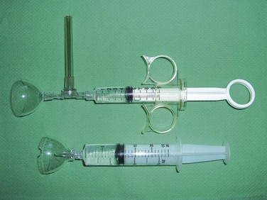
After the wound is anesthetized, cleansing of the wound should be performed by irrigation with normal saline at 8 to 12 psi of pressure. This can be accomplished by attaching an 18-gauge angiocatheter or a commercially available splash shield to a 20- or 30-mL syringe. At least 200 mL of irrigation is recommended. Irrijet and Zerowet were superior to an angiocatheter in preventing splatter during wound model irrigation.3 Zerowet was particularly effective in preventing splatter onto the irrigator’s face (Figure 7-1).3
Stay updated, free articles. Join our Telegram channel

Full access? Get Clinical Tree


