© Springer-Verlag Berlin Heidelberg 2017
George C. Velmahos, Elias Degiannis and Dietrich Doll (eds.)Penetrating Trauma10.1007/978-3-662-49859-0_1212. Intensive Care: Principles and Therapy
(1)
Division of Trauma/Critical Care and Emergency Surgery, University of Arizona, 1501 N Campbell Avenue, Rm 5411, 245063, Tucson, AZ 85724-5063, USA
Critical care management principles continue to be an extremely important aspect in the care of penetrating trauma patients, especially with the development of new technologies and advancing insight into human physiology. With the continual progression of new and exciting trauma research, the management of penetrating trauma necessitates all those involved in patient care be as current as possible in order to provide optimal care. The goal of this chapter is to provide a summary of important critical care principles allowing the reader to be successful in the management of penetrating trauma patients.
12.1 Metabolic Response to Trauma
The body’s response to a traumatic or surgical insult is basically the same. This involves an activation of the sympathetic nervous system and an increase in circulating catecholamines. Furthermore, endocrine stress hormones are released from the pituitary gland, as well as changes in the immune system including production of inflammatory cytokines in addition to a leukocytosis.
These neurohormonal changes cause tachycardia and fever, which taken together with tachypnea and leukocytosis form the systemic inflammatory response syndrome (SIRS). Metabolic changes lead to the retention of sodium and water, in addition to hyperglycemia that is compounded by insulin resistance, proteolysis of skeletal muscle, lipolysis of fat stores, and cytokine release, all of which contribute to a catabolic state.
It is presumed that this proinflammatory response is of overall benefit to the body as it responds to the insult and aids in recovery. However, in some patients, the degree of trauma is so great that despite adequate resuscitation, it overwhelms the capacity for recovery, and severe SIRS develops. A second “activating” event may also occur after the initial injury, which leads to a rapid downward spiral into multiorgan failure. The majority of modern critical care is designed to decrease the likelihood of this second event and/or to abrogate its effects on the patient.
12.2 ICU Monitoring
As critical care medicine continues to evolve and become more sophisticated, so do the means by which we monitor the critically ill. There continues to be a trend for noninvasive or minimally invasive monitoring for patients in the ICU. Numerous new devices are emerging which aide the clinician in making real-time decisions about the status and direction of patient physiology.
Arterial catheterization is still standard practice in most ICUs, not only to monitor arterial blood pressure but also to facilitate blood gas monitoring in ventilated patients. Complication rates are low, but thrombosis, pseudoaneurysm formation, and infection may still occur. The radial or femoral routes are preferred for ease of placement and minimal degree of complications. There are now several types of monitors that can be directly attached to the arterial line, giving real-time estimates of the cardiac output and index, stroke volume, systemic vascular resistance, etc.
Pulmonary arterial (PA) catheters continue to remain unfavorable in the ICU setting given their perceived high complication rate. We do believe however that there is still a small and specific patient population that will benefit from their use. This patient population includes those who are refractory to adequate volume resuscitation without obvious cause or who have cardiac dysfunction, either acute or chronic, where monitoring of cardiac output is necessary to guide clinical care.
Point-of-care ultrasonography (POCUS) has become extremely popular as a monitoring modality in the ICU over the past few years and is relatively inexpensive. It is a skill set that is now being taught routinely in both surgical residencies and critical care fellowships. The diagnostic impact of POCUS has been shown to be as high as 85 % whether that be confirming an expected diagnosis or resulting in a change of diagnosis and therefore a change in patient management. The ability to observe directly the heart, lungs, inferior vena cava, aorta, etc., with the ultrasound at bedside greatly improves the decision-making ability of the clinician, especially from a hemodynamics standpoint. Furthermore, ultrasonography has allowed for improvements in bedside procedures resulting in a lower complication rate, especially when it comes to the cannulation of vascular structures. Transesophageal ultrasonography has also gained popularity in the monitoring of volume status; however, this modality is more invasive and requires the patient to be intubated. Although the disadvantage of ultrasonography is that it is operator dependent, the application of this technology is vast and has demonstrated both reliability and reproducibility.
12.3 Neurological System, Pain Control, and Traumatic Brain Injury
One of the biggest challenges in the ICU is appropriately managing pain and agitation in critically ill patients. There are a number of considerations that you must take into account when choosing medications, not least of which are comorbidities that may affect the pharmacokinetics of the various agents. The Society of Critical Care Medicine recently published an in-depth set of guidelines for the management of pain, agitation, and delirium. Although pain and sedation management needs to be tailored to the individual patient, these guidelines have shown better outcomes for all critically ill patients and should be implemented in all ICUs:
- 1.
Optimize pain management first. This will allow for the use of less sedation and reduce the development of delirium. It is always preferable to use short-acting intravenous narcotic analgesics as infusions for initial pain control. Furthermore, early transition of patients to oral analgesia (e.g., oxycodone) via the enteral route is recommended as soon as the GI tract is available for use.
- 2.
Make light sedation the norm. Patients should be sedated to the point of comfort, especially if they are on the ventilator but should be alert enough to participate in their care. Oversedation only worsens the potential for delirium and can lengthen ventilation and ICU days. No matter which sedation medication is used, a sedation scale should be implemented with a specific target range so the patient does not become over sedated.
- 3.
Move away from routinely using benzodiazepines, especially in ICU patients who are at risk for or those who already have delirium. The choice of a benzodiazepine or other agents such as propofol as a sedative depends on the preference of the treating physician, as well as the patient’s hemodynamic stability. Both propofol and dexmedetomidine infusions, although short acting and more preferred than benzodiazepines, have deleterious effects on blood pressure requiring care when utilizing. Conversely, benzodiazepine infusions have long half-lives and should be avoided for maintenance sedation over many days.
- 4.
Implement effective delirium prevention and treatment strategies, using both nonpharmacologic and pharmacologic approaches. The development of delirium worsens overall mortality and should be avoided if possible. All ventilated patients should undergo daily “sedation vacations” to allow medication to wear off, which will allow more accurate assessment of the patient’s level of consciousness and further decrease delirium development. Regular reorientation, sunlight, providing familiarity in the patient room, appropriate sleep patterns, timely extubation, and early ambulation all assist in helping prevent delirium.
- 5.
Use antipsychotics judiciously. The more medications a patient receives, the more it can cloud their senses and judgment. Avoid additional medications, especially ones that affect the brain, as much as possible.
Traumatic brain injury is another major problem often seen in the ICU. Historically, the prognosis of most penetrating brain injuries was usually poor, especially if it is due to a missile that crosses the midline, resulting in the utilization of only a few resources to save the patient. Recent literature, however, has taken a more aggressive approach to patients with penetrating traumatic brain injury, especially those patients experiencing gunshot wounds to the head. Aggressive resuscitation with blood products and hypertonic saline has been shown to be independently associated with improvement in survival rates, even for those patients suffering bi-hemispheric missile trauma. The use of these aggressive resuscitative measures combined with rapid correction of coagulopathy as well as essential collaboration between the trauma, neurosurgery, emergency medicine, and nursing services has resulted in increased survival rates for gunshot wounds to the head from 10 to 46 %. Furthermore, the use of thyroid hormonal replacement therapy (the “T4 protocol,” which comprises of 1 ampule of 50 % dextrose, 2 g of methylprednisolone, 20 U regular insulin, and 20 mg levothyroxine) has been shown to significantly improve rates of organ procurement from those patients with fatal gunshot wounds to the head. With new literature showing increasing survival and organ procurement rates, the bias of resource use can no longer be used to preclude trauma surgeons from abandoning aggressive attempts to save patients with gunshot wounds to the brain.
12.4 Respiratory Failure, Acute Lung Injury, and ARDS
Many trauma patients suffer respiratory failure following injury and need to be maintained on invasive ventilation. Daily sedation vacations and spontaneous breathing trials have both been shown to facilitate earlier extubation. Weaning protocols that directly involve the nursing staff and respiratory therapists are highly effective. Computer-driven weaning protocols have also been developed and may be directly incorporated into ventilators in the future. Extubating patients as soon as possible is extremely important as it minimizes the risk of ventilator-associated pneumonia, one of the more serious nosocomial infections that patients develop in the ICU. These infections are associated with significant morbidity, mortality, and cost. Noninvasive ventilation may also have a role either as a means to avoid intubation while still providing ventilatory support or as a bridge following extubation to allow more time for the respiratory function to improve.
Acute respiratory distress syndrome (ARDS) is defined as acute hypoxemic respiratory failure with bilateral pulmonary infiltrates that is associated with both pulmonary and non-pulmonary risk factors. There are two main processes that contribute significantly to the development of ARDS: high permeability pulmonary edema and alveolar instability from the repetitive expansion and collapse of alveoli with tidal ventilation causing atelectrauma. In recent years, the definition of ARDS has changed to exclude the term “acute lung injury.” It is now referred to as “mild” ARDS. Furthermore, ARDS has been subcategorized into “moderate” and “severe” defined by the PaO2:FiO2 (200–300, 100–200, and <100, respectively).
As we have come to better understand the physiology behind ARDS, multiple modalities have been developed to better manage these patients. Prevention of ARDS development is most important through more judicious intravenous fluid resuscitation with better monitoring and the use of a “low tidal volume” strategy for ventilation. When ARDS does develop, airway pressure release ventilation (APRV) appears to be effective in reversing atelectasis and improving oxygenation, without requiring the deep sedation or paralysis that is usually necessary in the other forms of ventilation. Although high-frequency oscillatory ventilation has recently been shown to be detrimental as a rescue strategy for severe ARDS, inhaled nitric oxide or even extracorporeal membrane oxygenation has been shown to be successful in the right patient population. Furthermore, a recent study showed a 16 % decrease in mortality for those patients who develop severe ARDS who are placed in the prone position. Although proning a patient is not always easy, it requires minimal resources that should allow it to be performed in most ICUs.
12.5 Cardiac Failure
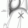
Cardiogenic shock is a relatively rare complication following penetrating trauma and is usually due to direct lacerations of the heart itself or secondary to underlying baseline cardiac disease. In cases of direct trauma to the heart, take care to avoid damage to the coronary arteries and veins if at all possible during surgical repair, as this will lead to infarction of the cardiac muscle distal to the injury. Pay attention to the possibility of damage to the cardiac valves, papillary muscles, and septae following penetrating injury, which is best evaluated by echocardiography, either via the transesophageal route during the initial operation or via the transthoracic method once the patient is in the ICU. It is recommended to routinely perform echocardiography postoperatively following cardiac stab wounds, and this should be done urgently if there are signs of cardiac dysfunction.
It is very common for trauma surgeons to care for a high volume of elderly patients who have multiple cardiac comorbidities such as coronary artery disease, valvular problems, or arrhythmias. Patients frequently have stents in place and are on clopidogrel and aspirin or are anticoagulated with warfarin, apixaban, dabigatran, or rivaroxaban. The risk of thromboses in these patients needs to be carefully weighed against the risk of ongoing hemorrhage if the anticoagulation is maintained. Furthermore, if there is ongoing bleeding, these medications may need to be reversed. Unfortunately, for many of these newer agents, reversal can be very challenging as there are not many agents to reverse them, and the agents that are currently available are very expensive. The reversal of warfarin has been fairly consistent for years utilizing vitamin K and fresh frozen plasma, although the prothrombin complex concentrates have recently become available and are more effective in reversing the effect of warfarin. However, for the factor Xa inhibitors (apixaban, rivaroxaban), prothrombin complex concentrate (PCC) is routinely used but not always successful. The thrombin inhibitors (dabigatran) can undergo hemodialysis to remove the effects of the anticoagulant, but the factor Xa inhibitors are resistant to hemodialysis.
The choice of vasopressor in cardiogenic shock will depend on the exact cause of the shock, dopamine being most beneficial in those patients needing inotropic support that are not tachycardic. Dobutamine is a better choice for patients with a history of congestive cardiac failure. Rarely, agents such as epinephrine or milrinone are utilized for right heart failure. In the most severe cases of cardiogenic shock, intra-aortic balloon pump placement may be required to maintain left ventricular function. Patients who develop cardiogenic shock should strongly be considered for placement of a pulmonary artery catheter as this can greatly help guide the clinical management.
Related posts:

Stay updated, free articles. Join our Telegram channel

Full access? Get Clinical Tree


