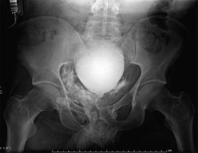
Fig. 20.1
(a) Grade IV right kidney injury with retroperitoneal hematoma (zone II) after multiple gunshot injuries to the back. IV contrast-enhanced CT scan of the abdomen is highly sensitive in diagnosing parenchymal injuries to the kidneys as well as in detecting active extravasation and associated intra-abdominal injuries. (b) Early contrast-enhanced abdominal CT scan after stab wound to the left flank. There is no contrast enhancement of the left kidney due to a renal arterial lesion
Initially, an early phase intravenous contrast CT scan of the abdomen and pelvis is performed. This investigation is highly sensitive in diagnosing parenchymal or vascular injuries to the kidneys as well as in detecting associated injuries (Fig. 20.1). To fully evaluate the collection system, a second CT scan is performed, approximately 10 min after intravenous contrast injection. This technique is known as CT intravenous pyelography (CT-IVP). These delayed-phase images are highly sensitive in diagnosing parenchymal injuries and proximal urine leaks or urinomas and in confirming bilateral functional renal moieties (Fig. 20.2). However, with a low sensitivity of 37 % to detect subtle ureteral injuries, small leaks at this location might be missed. Nevertheless, failure of the distal ureter to opacify on a contrast-enhanced CT scan should raise concern of an injury and should lead to further investigations or intraoperative evaluation of the affected ureter.
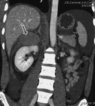
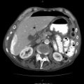
Fig. 20.2
Urinoma (white arrow) detected on IV contrast-enhanced CT scan after a stab wound to the right upper quadrant. The knife went through the right portion of the liver into the right kidney, where it perforated the major collection system
To further improve the value of the initial CT workup and to save time, a CT cystogram can be easily done simultaneously. CT cystography is equally as sensitive as conventional cystography for the detection of bladder rupture. Immediately before the CT scan, the urinary bladder is gently filled with approximately 350 ml of diluted iodine contrast through the urethral catheter. In the absence of a urethral injury, this procedure is safe and provides an accurate visual assessment of the integrity of the bladder. A urethral injury should be suspected in patients with pelvic fractures or penetrating injuries to the perineum. In these cases, you should consider a retrograde urethrogram before placing a urethral catheter.
20.3 Pyelography
Pyelography can be performed either as an excretory pyelography or as a retrograde pyelography. With a sensitivity of approximately 30 %, excretory pyelography is relatively insensitive for the diagnosis of renal injuries and urine leaks. However, in initially unstable patients, excretory pyelography may be useful to investigate the urinary tract intraoperatively after completion of the damage control procedure (Fig. 20.3). In the presence of hematuria or a suspicious penetrating injury tract, a “single shot” excretory IVP is performed intraoperatively. Ten minutes after intravenous injection of 2 cc/kg of contrast, a single abdominal plain film is taken. This investigation has been shown to obviate renal and ureteral exploration in 32 %.
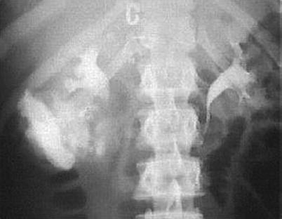
Fig. 20.3
Intraoperative excretory pyelography with complete proximal ureteral disruption. This intraoperative investigation of the urinary tract was made after completion of the damage control procedure
A retrograde pyelography is extremely sensitive in identifying ureteral injuries. However, in the emergency setting of patients sustaining abdominal penetrating injury, its value and practicability are limited. As an adjunct to CT-IVP or to confirm and further delineate the extent of a ureteral injury postoperatively, it is very helpful. Additionally, this investigation should be considered when planning further secondary surgical management of urinary tract injuries.
20.4 Cystography
All patients with abdominal gunshot wounds or pelvic fractures are at risk of having a ruptured bladder. Gross hematuria is very common in case of a bladder injury, occurring in over 95 % of cases. Imaging of the bladder using only excreted contrast material by CT scan or by conventional radiography is not adequate and has been shown to result in false-negative studies. The only definitive study to rule out a ruptured bladder is a retrograde static cystogram. The bladder is gently filled with approximately 350 ml of diluted iodine contrast through a urethral catheter. After that, an extravasation of contrast is captured by conventional plain film or, as described previously, by CT scan. In patients with a suspected urethral injury, a retrograde urethrogram should be considered before placing a urethral catheter. If a urethral rupture is found, the bladder is filled through a suprapubic tube.
20.5 Retrograde Urethrogram
The most common clinical finding in patients with urethral injuries are gross hematuria or blood at the meatus. Retrograde injection of contrast medium into the urethra is safe and has a high sensitivity for making the diagnosis of urethral rupture (Fig. 20.4). Multiple techniques have been described in the literature. We have had good results by inserting a 14-Fr Foley catheter at the meatus for about 3–4 cm to the fossa navicularis where the balloon is then gently inflated with 2–3 cc of sterile water. A Toomey syringe is then used to administer 30–40 cc or water-soluble contrast, and a plain conventional film is obtained, while the last 10 cc is instilled. A large extravasation without filling of the bladder indicates a complete disruption, whereas partial filling of the bladder with some extravasation is indicative of partial disruption of the urethra. If there is no extravasation, the catheter should be advanced into the bladder, and a cystogram should be added.