Immunofluorescence mapping is based on the detection of structural proteins of keratinocytes or of the dermo-epidermal junction using specific poly- or monoclonal antibodies. Through this method, the level of split formation can be determined by investigating the location of a given antigen in a natural or induced blister. This method also allows testing for the normal expression, reduction or absence of various structural proteins depending on the antibodies used. It has widely replaced transmission electron microscopy and is used as the initial laboratory test to prove the clinical diagnosis of epidermolysis bullosa.
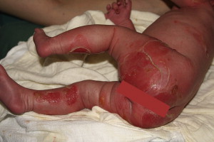
What is the diagnosis for a baby born with severe blisters and erosions ( Fig. 1 )? With an uninformative medical and family history, an experienced dermatologist considers an infectious, immunobullous, traumatic, or less frequently, an inherited disorder. The spectrum of differential diagnoses is extensive, and distinction can be difficult. Diagnostic routine tests such as microbial testing should be performed first, frequently followed by skin biopsy for routine histologic, immunohistochemical and, sometimes, ultrastructural assessment. In epidermolysis bullosa (EB), microscopic results first show an unremarkable routine histology, except for a split formation in distinct layers of the skin and a negative routine immunohistochemistry. As the newborn develops more blisters every day, especially on sites of mechanical trauma, the diagnosis of inherited EB has to be considered.
The challenges in the diagnosis of EB are identical worldwide. To achieve a correct diagnosis and classification of the disease, the first laboratory tool to be used successfully was transmission electron microscopy (TEM), and this has been the gold standard laboratory test for many years (see the article by Eady and Dopping-Hepenstal elsewhere in this issue for further exploration of this topic). However, TEM is time-consuming and expensive, and the results are, to a high degree, operator-dependent and sometimes inaccurate. In addition, there are only a few laboratories with appropriate experience and skills to analyze and interpret EB samples in TEM. In 1981, the authors described the method of immunofluorescence (antigen) mapping (IFM), which is based on the detection of structural proteins of keratinocytes or of the dermo-epidermal junction using specific poly- or monoclonal antibodies. Using this method, the level of split formation can be determined by investigating the location of a given antigen (eg, type 4 collagen) in a natural or induced blister. This method also allows testing for the normal expression, reduction or absence of various structural proteins depending on the antibodies used.
Based on the localization of the level of blistering in the skin as determined by immunohistochemistry and/or TEM, the hereditary mechanobullous diseases are classified into 3 major groups: EB simplex (EBS), junctional EB (JEB), and dystrophic EB (DEB). A fourth group, the Kindler syndrome, was added according to the new classification published in 2008.
There are various poly- or monoclonal antibodies that recognize structural proteins of keratinocytes or of the dermo-epidermal basement membrane zone (BMZ), which is known to be involved in the pathogenesis of EB. Fortunately, these antibodies are available worldwide for the frequently used IFM. IFM has widely replaced TEM and is now used as the first laboratory test to prove the clinical diagnosis of EB and to distinguish subtypes of EB. It is also the base for distinguishing the target proteins for mutation analysis.
In this article, IFM as a crucial diagnostic tool is described and the possibility of a worldwide cooperation in the diagnosis of EB is discussed.
Materials and methods
Biopsy
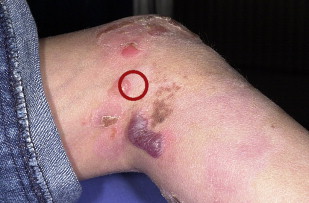
The method for taking a biopsy for EB is reviewed elsewhere in this issue (see the article by Intong and Murrell elsewhere in this issue for further exploration of this topic). A biopsy of patient skin, including parts of a fresh blister and of normal-appearing skin ( Fig. 2 ), facilitates the determination of the split formation and allows conclusions about the major types of EB.
Transport to the Specialized Laboratory
The biopsy sample should be immediately placed into Michel medium as originally described by Michel and colleagues in 1973 and modified by Vaughan and colleagues in 1995: 1 M citrate buffer pH 7.4, 2.5 mL; 0.1 M magnesium sulfate, 5 mL; 0.1 M N-ethylmaleimide, 5 mL; ammonium sulfate 55 g; distilled water, 87.5 mL; total volume 100 mL adjusted to pH 7.4 with 1 M sodium hydroxide. In this medium, the samples can be stored up to 28 days at room temperature and sent worldwide to any specialized laboratory that performs IFM. On receipt, the sample has to be rinsed immediately in phosphate buffered saline (PBS) for a few hours to reduce background staining and improve diagnostic sensitivity. Then the sample can be processed by cutting cryostat sections or snap frozen in liquid nitrogen or stored in a plastic tube without any liquid in a freezer at −20°C (or better, at −80°C) until further processing.
In a recent cooperation between an EB group in Mexico and the EB House at the Department of Dermatology in Salzburg, Austria, 48 biopsy samples were sent from Mexico to Austria. It was confirmed that the above-mentioned conservation and transport methods allow an excellent IFM quality leading to an appropriate classification (manuscript in preparation).
Antibodies
The primary antibodies for IFM are generated from different animal sources and bind to specific structural proteins of the skin that are relevant in the pathogenesis of various types of EB ( Table 1 ). According to the proteins targeted for mutation analysis, we use IgG antibodies against cytokeratin 5, cytokeratin 14, plectin, α6 integrin, β4 integrin, type 17 collagen (180 kDa BPAG2), laminin 332, and type 7 collagen. We also use anti-type 4 collagen (present in the lamina densa of the dermo-epidermal BMZ) antibodies for better visualization of the level of split formation, especially in patients with DEB. All primary antibodies are IgG class, and most of them are monoclonal, raised in mice. Therefore, the second antibody has to be a mouse-specific anti-IgG antibody, usually conjugated to the fluorescent dye, fluorescein isothiocyanate (FITC). An exception is the antibody against α6 integrin where rat is the source, and thus the second antibody has to be anti-rat IgG. The FITC bound to the second antibody produces a specific fluorescent signal at 450 to 490 nm and allows the visualization of specific antibody binding under the IF microscope. Depending on the frequency of requested IFMs and the final dilution necessary for IFM, different amounts of the original primary and secondary antibodies should be divided into aliquots and kept in the refrigerator until expiry date given on the bottle of the stock solution. Working dilutions are always prepared fresh from these aliquots and are not reused.
| 1st Antibodies | Dilution | Host | Company | Catalog Number | EB Subtype |
|---|---|---|---|---|---|
| Cytokeratin 5 | 1:50 | Mouse | Millipore | MAB 3224 | EBS |
| Cytokeratin 14 | 1:100 | Mouse | Millipore | MAB 3232 | EBS |
| Plectin (5B3) | 1:2 | Mouse | Wiche (personal communication) | EBS, JEB | |
| α6 integrin | 1:50 | Rat | Millipore | MAB 1378 | EBS, JEB |
| β4 integrin | 1:50 | Mouse | Millipore | MAB 1964 | EBS, JEB |
| Collagen 17 | 1:20 | Mouse | In-house (EB laboratory) | JEB | |
| Laminin 332 a | 1:50 | Mouse | Millipore | MAB 1949 | JEB |
| Collagen 4 | 1:50 | Mouse | Millipore | MAB 3326 | |
| Collagen 7 | 1:50 | Mouse | Millipore | MAB 1345 | DEB |
| 2nd Antibodies | |||||
| IgG | 1:100 | Goat anti-mouse | Millipore | AP 124F | |
| IgG | 1:100–400 | Rabbit anti-rat | Dako | F 0234 | |
Related posts:
 How to Take Skin Biopsies for Epidermolysis Bullosa
 Wound Management for Children with Epidermolysis Bullosa
 Genitourinary Tract Involvement in Epidermolysis Bullosa
Growth and Pubertal Delay in Patients with Epidermolysis Bullosa
Genitourinary Tract Involvement in Epidermolysis Bullosa
Growth and Pubertal Delay in Patients with Epidermolysis Bullosa
 Epidermolysis Bullosa Care in Germany
Epidermolysis Bullosa Care in Israel
Epidermolysis Bullosa Care in Germany
Epidermolysis Bullosa Care in Israel
Stay updated, free articles. Join our Telegram channel

Full access? Get Clinical Tree


