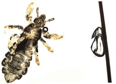
Fig. 16.2Head louse (Pediculus humanus capitis) and an empty egg casing (the operculum is at the top) attached to a hair shaft.
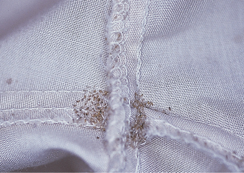
Fig. 16.3Body lice eggs in the seams of clothing.Courtesy, Yale Dermatology Residents’ Slide Collection. From Bolognia JL, Jorizzo JL, Schaffer JV. Dermatology, 3e. London: Saunders, 2012, with permission.
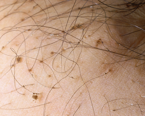
Fig. 16.4Crab lice and eggs on pubic hair.Courtesy, Louis A Fragola, MD. From Bolognia JL, Jorizzo JL, Schaffer JV. Dermatology, 3e. London: Saunders, 2012, with permission.
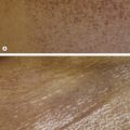
Fig. 16.5Range of cutaneous lesions in scabies. Crusted scabies may show prominent hyperkeratosis of acral sites.From Bolognia JL, Schaffer JV, Duncan KO, Ko CJ. Dermatology Essentials, 1e. Philadelphia: Saunders, 2014, with permission.
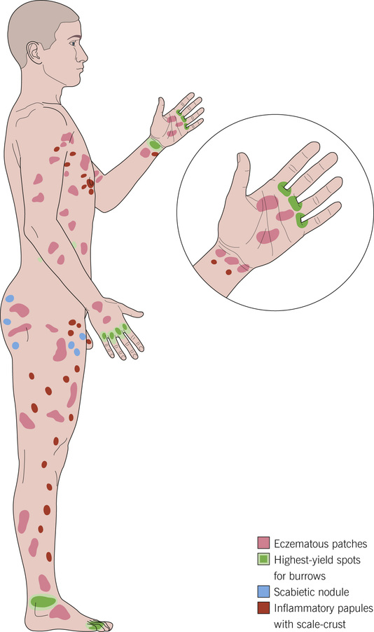
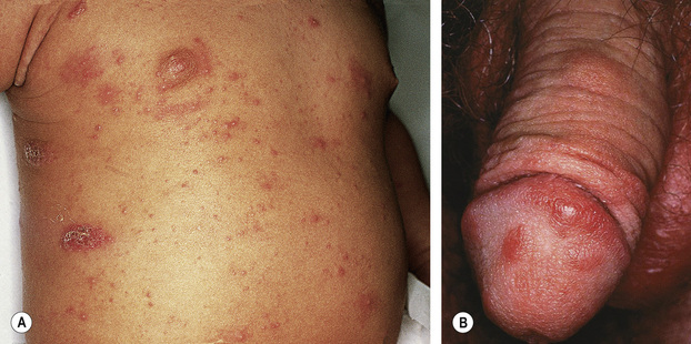
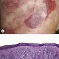
Fig. 16.6Scabies infestation. A Eczematous patches and inflammatory papules. B Scabietic nodules.A, Courtesy, Yale Dermatology Residents’ Slide Collection. B, Courtesy, Robert Hartman, MD. A, From Bolognia JL, Jorizzo JL, Schaffer JV. Dermatology, 3e. London: Saunders, 2012, with permission. B, From Bolognia JL, Schaffer JV, Duncan KO, Ko CJ. Dermatology Essentials, 1e. Philadelphia: Saunders, 2014, with permission.
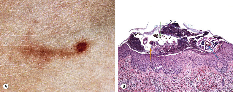
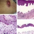
Fig. 16.7Scabies. A Scabies linear burrow. B Microscopic mite (blue arrow), scybala (green arrow), and egg casings (orange arrow).A, Courtesy, NYU Slide Collection. A, From Bolognia JL, Jorizzo JL, Schaffer JV. Dermatology, 3e. London: Saunders, 2012, with permission.
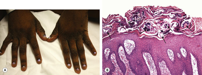
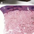
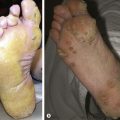
Fig. 16.8Crusted scabies. The hyperkeratotic lesions, most typically on the hands, house numerous mites (arrows) and scybala (*).Courtesy, Yale Dermatology Residents’ Slide Collection.
Demodex Folliculitis
Due to Demodex spp.
Erythematous papules and/or pustules on the face (Figs 16.9,16.10)
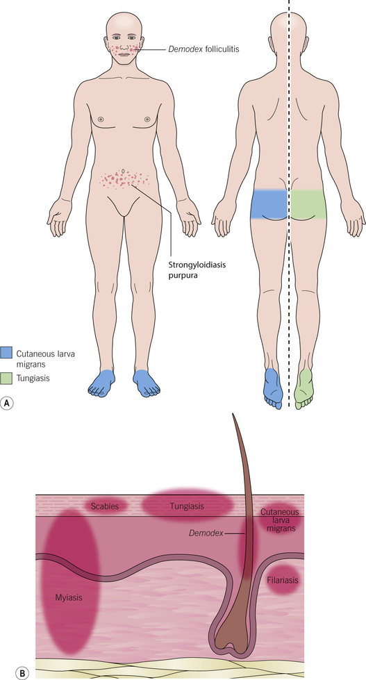
Fig. 16.9Distribution of selected infestations. A Typical sites affected in Demodex folliculitis, strongyloidiasis, tungiasis, and cutaneous larva migrans. B Histopathologic location of selected arthropods/helminths. Microfilariae of filariasis are located in the dermis, but adult worms are in subcutaneous tissue or lymphatics.
Only gold members can continue reading. Log In or Register to continue