Describe the anatomy of the branchial apparatus (see Fig. 49-1).
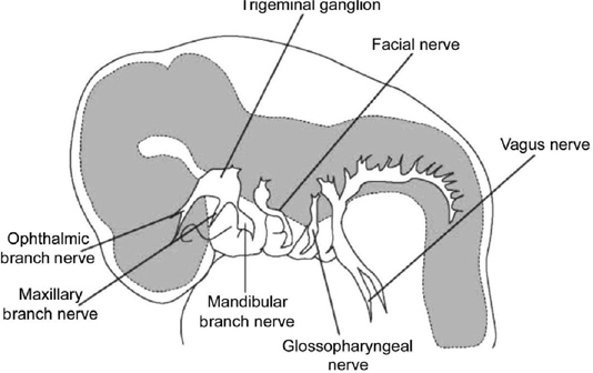
Figure 49-1 Branchial arches.
The branchial apparatus consists of five paired mesodermal arches that are separated by invaginations of ectoderm and endoderm known, respectively, as clefts and pouches. Each arch has a cartilage bar, an artery, and a nerve. The derivatives of the cartilage of each arch form the facial skeleton and laryngeal framework, while the artery and nerve supply and innervate the derivatives of the arch.
 What is derived from the ectoderm/branchial clefts?
The ectoderm of the first cleft forms the major salivary glands, the mucosa of the oral cavity, and the lining of the anterior two-thirds of the tongue, while the ectoderm of the second, third, and fourth arches fuse to form a common cervical sinus (of His), which normally degenerates.
 What is derived from the endoderm/branchial pouches?
The endoderm of the branchial pouches forms the middle ear, the glandular structures of the oropharynx, the parathyroid glands, and the thymus.
 What is the nerve of the first branchial arch?
The trigeminal nerve.
 What is the artery of the first branchial arch?
The artery of the first arch partly degenerates and partly remains as the maxillary artery.
 What are the derivatives of the cartilage of the first arch?
Meckel’s cartilage is the cartilage of the first arch and its derivatives are the mandible, the malleus (except for the manubrium), and the incus (except for the long process).
 What are the muscular derivatives of the first branchial arch?
The muscular derivatives are the muscles of mastication (temporalis, masseter, medial and lateral pterygoids), the tensor tympani, the mylohyoid, anterior belly of the digastric, and the tensor veli palatini.
 What is the nerve of the second branchial arch?
The facial nerve.
 What is the artery of the second branchial arch?
The stapedial artery, which usually degenerates in normal development but occasionally persists in adulthood.
 What are the derivatives of the cartilage of the second arch?
Reichert’s cartilage is the cartilage of the second arch and its derivatives are the manubrium of the malleus, the long process of the incus, the stapes suprastructure, the styloid process, the stylohyoid ligament, and the body and lesser cornu of the hyoid bone.
 What are the muscular derivatives of the second branchial arch?
The muscular derivatives of the second arch are the muscles of facial expression, the platysma, stylohyoid, posterior belly of the digastric, and the stapedius muscle.
 What is the nerve of the third branchial arch?
The glossopharyngeal nerve.
 What is the artery of the third branchial arch?
The internal carotid artery.
 What are the derivatives of the cartilage of the third arch?
The body and greater cornu of the hyoid are the skeletal derivatives of the third arch.
 What are the muscular derivatives of the third branchial arch?
The stylopharyngeus is the only muscle of the third arch.
 What is the nerve of the fourth branchial arch?
The superior laryngeal nerve.
 What is the artery of the fourth branchial arch?
The aortic arch is derived from the left arch, and the right subclavian artery is derived from the right arch. If the right fourth arch artery degenerates, the right subclavian artery will arise from the dorsal aorta and run posterior to the esophagus (retroesophageal subclavian).
 What are the derivatives of the cartilage of the fourth arch?
The thyroid and cuneiform cartilage of the larynx.
 What are the mesodermal derivatives of the fourth branchial arch?
The inferior pharyngeal constrictor, cricopharyngeal, and cricothyroid are the muscular derivatives of the fourth arch.
 What is derived from the fifth branchial arch?
The fifth arch degenerates and has no derivatives in the human.
 What is the nerve of the sixth branchial arch?
The recurrent laryngeal nerve.
 What is the artery of the sixth branchial arch?
The pulmonary artery.
 What are the derivatives of the cartilage of the sixth arch?
The skeletal derivatives are the cricoid, arytenoids, and corniculate cartilage.
 What are the muscular derivatives of the sixth branchial arch?
The intrinsic muscles of the larynx.
 What are branchial cleft anomalies?
Related posts:
Stay updated, free articles. Join our Telegram channel

Full access? Get Clinical Tree


