EMBRYOLOGY 
General Organization of the Limb
 What tissues comprise the limb bud?
Mesenchyme covered by ectoderm. The limbs grow by proliferation of mesenchyme.
 Which genes regulate patterning in limb development?
Homeobox-containing (HOX) genes.
Timing
 When do the limb buds first appear?
Toward the end of the 4th week of development, at which time a group of mesenchymal cells in the lateral mesoderm are activated. The buds are visible by day 26 to day 27.
 When is the critical period for upper limb development?
Twenty-four to 36 days after fertilization.
 When are finger buds visible?
The end of week 6.
 When does ossification occur?
Between weeks 8 and 12, ossification of the cartilaginous framework occurs. Epiphyses gradually ossify until the termination of puberty.
 What are the last bones to ossify within their cartilaginous framework?
The carpal bones in which ossification begins during the first year of life.
 Does sensory or motor innervation occur first?
Sensory axons enter the limb after motor axons and use these for guidance.
 When is nervous system myelination completed?
Around 2 years of age.
Axes of Development
 What is responsible for proximodistal development?
The apical ectodermal ridge (AER), which expresses endogenous fibroblast growth factors.
 What is responsible for radioulnar development?
The zone of polarizing activity, found in the posterior margin of the limb bud, and activated by fibroblast growth factors from the AER that cause Sonic hedgehog (SHH) gene expression.
 What is responsible for dorsoventral development?
Expression of wingless-type MMTV integration site family member 7A WNT7 and engrailed-1 (EN1) from the dorsal and the ventral epidermis, respectively.
Tissue Differentiation
 What process is responsible for syndactyly occurrence?
Apoptosis, likely mediated by bone morphogenic proteins signaling TGF-β.
 How common is syndactyly and which digits are most commonly affected?
Occurs in 1 in 2,200 births, most commonly affecting the webspace between the middle and ring fingers.
 Which arteries supply the limb buds?
Dorsal intersegmental arteries arising from the aorta.
Congenital Deformities
 Outline the American Society for Surgery of the Hand classification of congenital deformities of the hand.
The original classification scheme was proposed by Swanson:
i. Failure of formation
ii. Failure of differentiation
iii. Duplication
iv. Overgrowth
v. Undergrowth
vi. Constriction band syndromes
vii. Generalized anomalies and syndromes
 HAND ANATOMY
Surface Anatomy
 What is the anatomic snuffbox?
The area bounded by the tendons of the abductor pollicis longus (APL) and extensor pollicis brevis (EPB) anteriorly and the tendon of the extendor pollicis longus posteriorly. The floor is formed by the scaphoid and trapezium, and it is a frequent site of tenderness after scaphoid fracture (see Fig. 29-1).
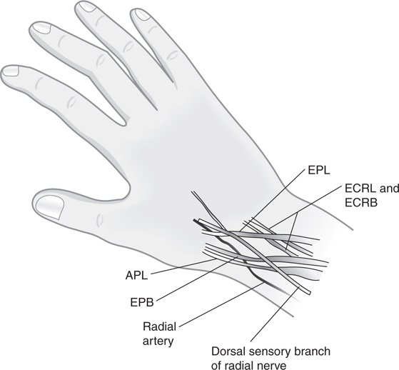
Figure 29-1 Diagram demonstrating boundaries of the anatomic snuffbox.
 Where is the metacarpophalangeal (MCP) joint relative to the MCP palmar flexion crease?
Proximal.
 What is the “fixed unit” of the hand?
The distal carpal row and second and third metacarpals.
Soft Tissue
Fingertip
 What is a felon?
A subcutaneous abscess of the distal digital pulp. It involves the septal compartments and causes compartment syndrome of the distal phalangeal pulp. If the pad is not involved, then it is an “apical infection” instead.
 What is the normal value for moving two-point discrimination of the fingertip?
Two to 3 mm, with best discrimination occurring in individuals in their 20s and in ulnar-sided digits. A score of 7 out of 10 correct answers determines the value of two-point discrimination.
Nail and Nail Bed
 What is the perionychium? Paronychium? Hyponychium? Eponychium? Lunula?
The perionychium includes the nail bed, nail fold, eponychium, paronychium, and hyponychium. The paronychium is the skin on either side of the nail bed. The hyponychium is skin distal to the nail bed. The eponychium is the skin proximal to the nail that covers the nail fold. The white arc in the proximal nail is the lunula and is the distal extent of the germinal matrix. Distal to this is the sterile matrix (see Fig. 29-2).
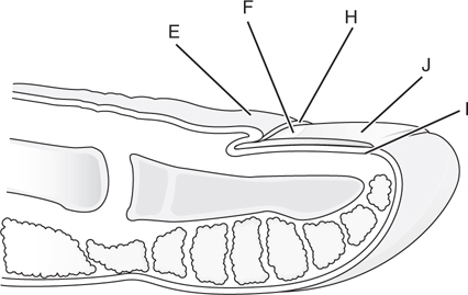
Figure 29-2 Perionychium (E), lunula (F), nail bed (G), germinal matrix (H), sterile matrix (I), nail plate (J), hyponychium (K), and distal groove (L).
 Which tissues contribute to nail growth?
The germinal matrix produces approximately 90% of the nail, while the sterile matrix adds a thin layer of cells underneath the nail to ensure its adherence.
 What is a paronychia?
Acute paronychia is infection of the paronychial tissues.
 How long does a nail take to grow?
Approximately, 6 months for the entire nail to regrow.
Muscles
 Name the median innervated intrinsic muscles of the hand.
The intrinsic muscles of the hand are innervated by the ulnar nerve, except the radial two lumbricals, opponens pollicis, abductor pollicis brevis, and superficial head of the flexor pollicis brevis (mnemonic: Meat-LOAF).
 Which muscle is most involved in lateral epicondylitis (“tennis elbow”)?
Extensor carpi radialis brevis (ECRB).
 What are the functions of the palmar and dorsal interossei?
The interossei originate from the metacarpal shafts and form the lateral bands, aiding in the function of the lumbrical muscles. In addition, the Palmar interossei ADduct the fingers and the Dorsal interossei ABduct the fingers (mnemonic: PAD-DAB).
 What is a lumbrical plus deformity?
A lumbrical plus deformity is characterized by paradoxical extension of the proximal interphalangeal (PIP) joint with attempted digital flexion. This is due to excessive tension on the lumbrical muscles through the FDP tendon with flexion causing pull on the lateral bands, often the result of FDP retraction after a distal amputation.
 Name the thenar muscles. What order are they in?
Superficial to deep, they are:
• abductor pollicis brevis
• flexor pollicis brevis
• opponens pollicis
• adductor pollicis
 Name the hypothenar muscles.
• Palmaris brevis
• Abductor digiti minimi
• Flexor digiti minimi brevis
• Opponens digiti minimi
 Which muscle group in the forearm is most susceptible to compartment syndrome?
The flexor digitorum profundus.
 Name the muscles of the mobile wad.
Brachioradialis, ECRB, extensor carpi radialis longus (ECRL).
 How can you test for extensor pollicis longus (EPL) function?
Ask the patient to rest their hand palm down on a table and lift the thumb—only the EPL can move lift the thumb dorsal to the plane of the palm.
Fascia and Ligaments
 What are Cleland’s and Grayson’s ligaments?
Cleland’s ligaments connect the digital fascia to the sides of the phalanges and are not involved in Dupuytren disease. They lie dorsal to the neurovascular bundle. Grayson’s ligaments connect the tendon sheath to the digital fascia and are often involved in Dupuytren disease. They lie volar to the neurovascular bundle.
 What are natatory ligaments?
This is another name for the superficial transverse metacarpal ligaments and these help to create the webspaces. These coalesce distally with the spiral bands and may be involved in Dupuytren’s disease.
 What are the boundaries and contents of the carpal tunnel?
1. Roof: transverse carpal ligament
2. Floor: carpal bones
3. Radial border: scaphoid and trapezium
4. Ulnar border: pisiform and hamate
5. Contents: the median nerve and nine tendons (flexor digitorum superficialis [FDS], flexor digitorum profundus [FDP], and flexor pollicis longus [FPL])
 What are the boundaries and contents of Guyon’s canal?
1. Roof: volar carpal ligament and pisohamate ligament
2. Floor: transverse carpal ligament
3. Ulnar wall: pisiform
4. Contents: ulnar nerve and artery
 What is the oblique retinacular ligament?
Related posts:
Stay updated, free articles. Join our Telegram channel

Full access? Get Clinical Tree


