Summary and Key Features
- •
Fibroblast secretome, also known as cell-conditioned medium, contains growth factors and exosomes and plays a major role in skin repair and regeneration.
- •
Topical application of fibroblast secretome alters expression of key extracellular matrix and skin health genes.
- •
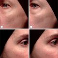
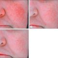
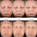
Clinical evidence shows increased collagen production and decreased facial fine lines and wrinkles.
- •
Growth factors and exosomes derived from different cells or different conditions have different biological activity.
- •
Growth factors and exosomes must be stabilized in formulations and may be combined with other modalities of skin rejuvenation such as lasers and topical antioxidants.
See .
Introduction
Exposure to environmental pollution and solar radiation causes cumulative damage that accelerates normal chronologic aging and exacerbates injury to skin tissue, resulting in photodamage. Consumer interest in correcting signs of photodamage such as wrinkles, dyspigmentation, sagging, and surface roughness is increasing as the population ages. Treatments include topical retinoids and antioxidants, chemical peels, dermabrasion, laser and other energy-based devices, and various lifting surgeries, depending on the severity of the damage.
Within the past decade, researchers have focused on the pathophysiology of photodamage and have found correlations with certain aspects of acute and chronic wound healing. Of specific interest are the effects of secreted components (secretome) from fibroblasts on the process of wound healing and skin repair. Fibroblast secretome contains growth factors, various peptides, and exosomes that mediate signaling pathways between and within cells. Exosomes contain peptides, small growth factors, and small DNA and RNA molecules in a submicron-sized particle made of cell membranes. After a wound has been inflicted, secretome from a variety of cells floods the wound site and interacts synergistically to initiate and coordinate each phase of wound healing. This process is complex and not completely understood. Many studies demonstrate the importance of individual growth factors in the repair of damaged tissue, but research into the phases of wound healing has demonstrated that it is the interaction of multiple growth factors, peptides, and RNAs that is vital to tissue regeneration. Advances in our understanding of the role of exosomes present in cellular secretome have created renewed interest in using secretome from various cells for prevention and correction of aging-related skin changes. Multiple clinical studies now show accelerated skin repair and regeneration after topical use of fibroblast secretome.
Photodamage Effects on Skin Tissue
Photodamage occurs predominantly within the epidermis and the upper papillary dermis. Histologic studies demonstrate that ultraviolet (UV) exposure disrupts the normal architecture of connective tissue within the dermis. The dermal extracellular matrix (ECM) is composed primarily of type I collagen, although type III collagen, elastin, proteoglycans, and fibronectin are also included in smaller amounts. UV exposure decreases collagen and elastin and alters the cross-linked structure of collagen and elastin fibers within the dermal ECM. Abnormal elastic material containing elastin and fibrillin accumulates and appears to occupy areas of lost collagen. This deposition of abnormal elastic material is called solar elastosis . Glycosaminoglycans (GAGs), a type of proteoglycan that is a component of the ECM, are polysaccharide molecules that bind to water, forming a hydrated, space-filling polymer between collagen and elastin fibers that helps support skin tissue. In photodamaged skin, GAGs are abnormally deposited in the elastotic tissue rather than between collagen and elastin fibers. The clinical result of decreased collagen and elastin and disruption of the normal support architecture is the appearance of wrinkles, sagging skin, uneven pigmentation, hyperpigmentation, and thickened or leathery skin texture. Although chronologically aging skin also develops wrinkles, photodamaged skin is differentiated histologically by the presence of solar elastosis.
Biochemical Pathways of Skin Aging
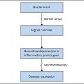
Fig. 16.1 shows a simplified summary of major pathways involved in the aging process. Absorption of solar radiation by chromophores in the skin as well as cellular oxidative metabolism results in the formation of reactive oxygen species (ROS). ROS increase oxidative phosphorylation of cell-surface receptors causing activation of the transcription factor activator protein-1 (AP-1) and nuclear factor-kappa B (NF-κB).
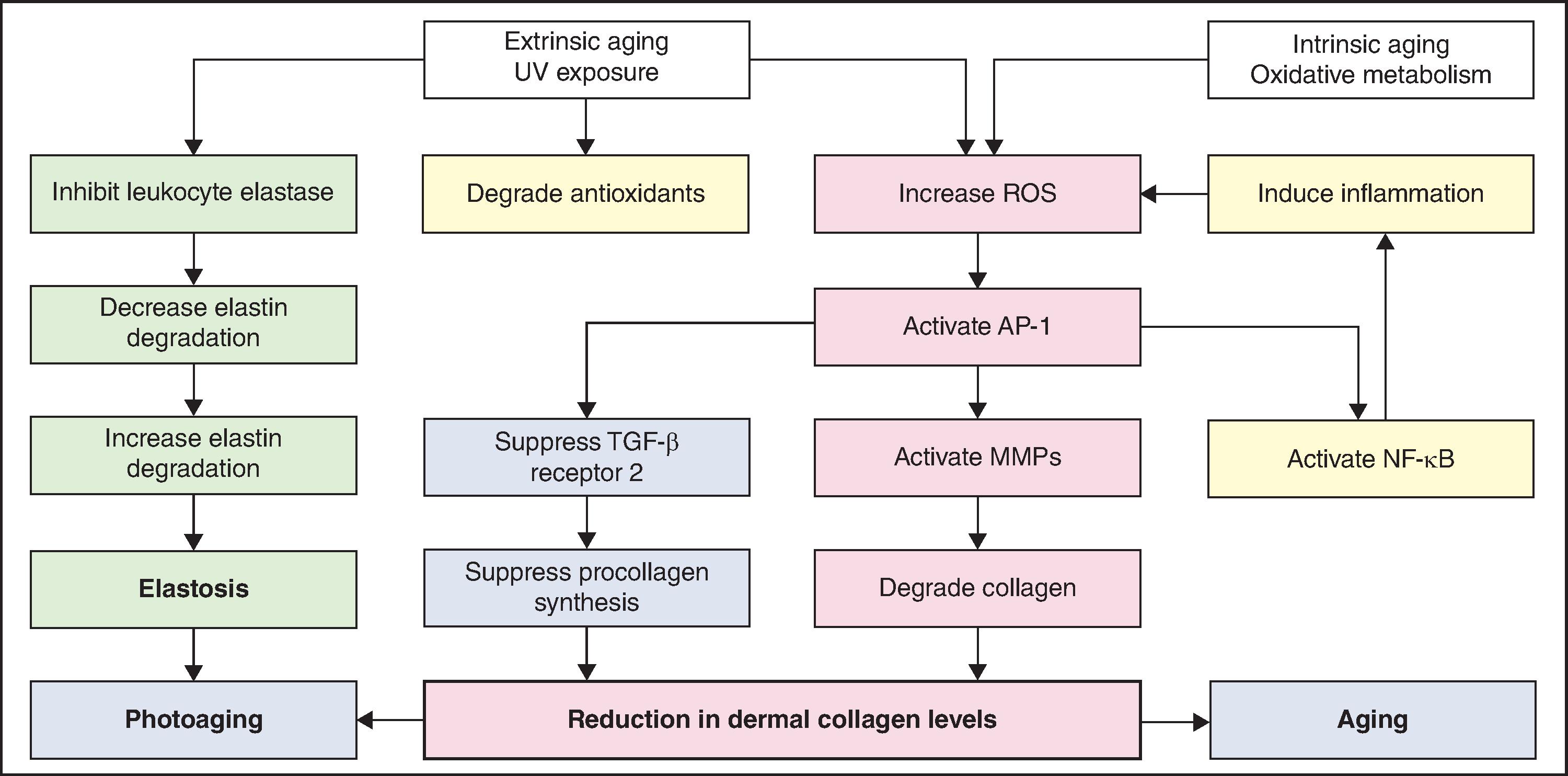
AP-1 stimulates transcription of matrix metalloproteinase (MMP) growth factor genes in fibroblasts and keratinocytes and inhibits type I procollagen gene expression in fibroblasts. MMPs produce degradation of fibrillar type I and type III collagen. Activity of MMP is decreased by binding with tissue inhibitors of metalloproteinase (TIMPs). ROS inactivate TIMPs, thereby increasing MMP activity. AP-1–mediated reduction in synthesis of procollagen appears to result from two mechanisms: interference of AP-1 with type I and type III procollagen gene transcription and blocking the profibrotic effects of transforming growth factor beta (TGF-β) by impairment of the TGF-β type 2 receptor/Smad pathway.
Activation of NF-κB stimulates transcription of proinflammatory cytokine genes including interleukin-1 (IL-1), tumor necrosis factor-alpha (TNF-α), IL-6, and IL-8. Inflammation resulting from these cytokines increases secretion of ROS and more cytokines, further enhancing the effect of UV exposure. Inflammation causes protease-mediated degradation of elastin and UV exposure causes formation of abnormal elastin by fibroblasts. UV light is also an inhibitor of leukocyte elastase, thereby increasing accumulation of elastotic materials. The accumulation of elastotic materials is accompanied by degeneration of the surrounding collagenous network.
The overall effects of these interlinked biochemical activities are reduction of procollagen synthesis, increase of collagen degradation in the dermal ECM, and increase in irregular elastin deposition.
Cell Secretome and Growth Factors in Skin Repair
Hundreds of growth factors and other regulatory molecules have been identified in skin. Those that are important in skin repair and wound healing include cytokines involved in immune response and phagocytosis, and growth factors that induce the synthesis of new collagen, elastin, and GAGs (components of the dermal ECM) that are abnormally affected by UV radiation. Table 16.1 lists skin-related biological activities of some of the most important proteins in cellular secretome. Additional research is needed to better understand the role of thousands of exosome components in skin repair and regeneration.
| Growth Factor or Cytokine | Skin-Related Biological Activity |
|---|---|
| EGF | Epidermal and dermal regeneration |
| TGF-β1 | Keratinocyte migration; chemotactic for macrophages and fibroblasts TGF-β1: Type III collagen formation TGF-β3: Type I collagen formation |
| TGF-β2 | |
| TGF-β3 | |
| FGF2/bFGF | Angiogenesis; activation of endothelial cellular proliferation and differentiation |
| FGF4 | Activation of cellular proliferation and differentiation |
| FGF6 | |
| FGF7/KGF | Keratinocyte proliferation; epithelial cell–specific growth factor |
| FGF9 | Hair follicle development after wound |
| VEGF | Influences vascular permeability and angiogenesis to improve tissue nutrition |
| PDGF AA | Chemotactic for macrophages, fibroblasts, macrophage activation, fibroblast mitogen, and matrix production |
| PDGF BB | |
| PGF | Promotes endothelial cell growth |
| HGF | Strong mitogenic activities; three-dimensional tissue regeneration and wound healing |
| IGF1 | Endothelial cell and fibroblast mitogen |
| GCSF | Colony-stimulating factors stimulate the development of neutrophils and macrophages |
| GM-CSF | |
| M-CSF | |
| CTGF | Cell adhesion, migration, proliferation, angiogenesis, and fibrosis |
| IL-1 | Early activator of growth factor expression |
| IL-2 | Enhances epithelial wound healing |
| IL-3, IL-4, IL-5 | Leukocyte maturation/degranulation during inflammatory phase |
| Il-6 | Mediator of acute phase response to wound with IL-1 |
| IL-7, IL-8, IL-15 | Leukocyte activation and proliferation during inflammatory phase |
| IL-9 | Keratinocyte proliferation and differentiation |
| IL-10 | Inhibits proinflammatory cytokines |
| IL-13 | Stimulates production of IL-6 |
| MMP1, MMP2 | Degrade collagen and other extracellular matrix components |
| MMP9 | Degrades type IV collagen |
| TIMP1, TIMP2 | Prevent enzymatic degradation of collagen and other extracellular matrix components |
| TNF | Induces inflammation to trigger wound healing |
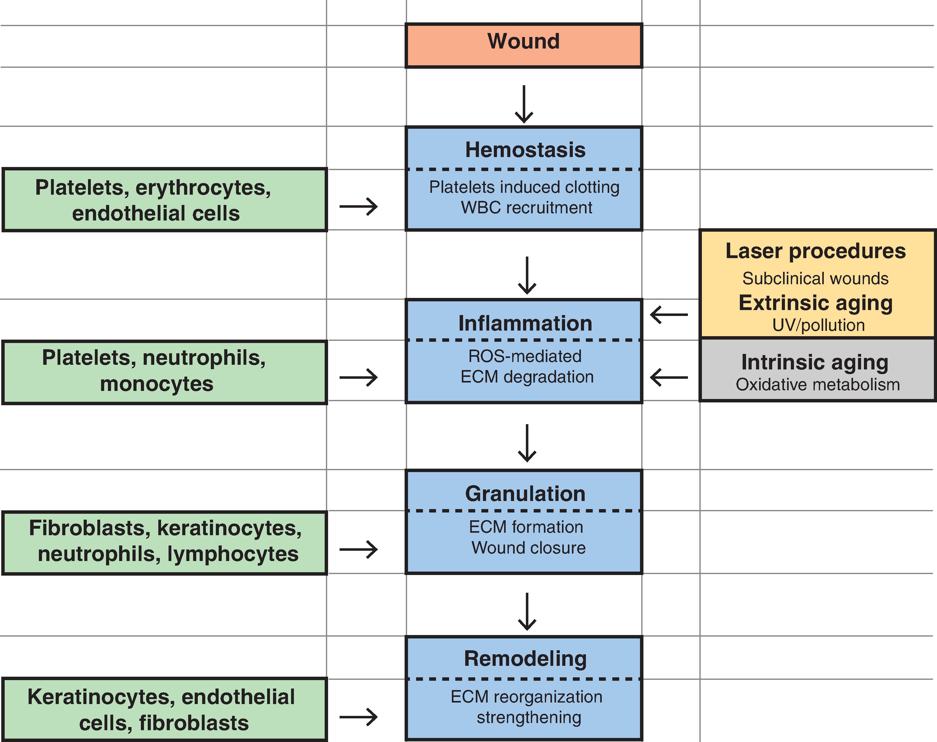
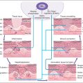
After injury, cytokines and other growth factors flood the wound site to mediate the inflammatory response, promote new cell growth, and decrease wound contraction and scarring. The process of wound healing is commonly divided into four overlapping phases that describe physiologic responses to injury. These phases include hemostasis, inflammation, proliferation, and remodeling. Fig. 16.2 summarizes each phase of wound healing along with the predominant cells involved. During hemostasis, platelets release various cytokines, growth factors, and exosomes at the wound site to promote chemotaxis and mitogenesis. In the inflammatory stage, neutrophils and monocytes migrate to the wound site to initiate phagocytosis and to release their secretome that attract fibroblasts. The proliferation phase is marked by epithelialization, angiogenesis, granular tissue formation, and collagen deposition. During proliferation, keratinocytes restore barrier function to the skin, and its secretome stimulates the expression of new keratin proteins. Fibroblasts produce collagen that is deposited in the wound bed. This cycle of collagen production and growth factor secretion continues in a type of autocrine feedback loop of continuous wound repair.