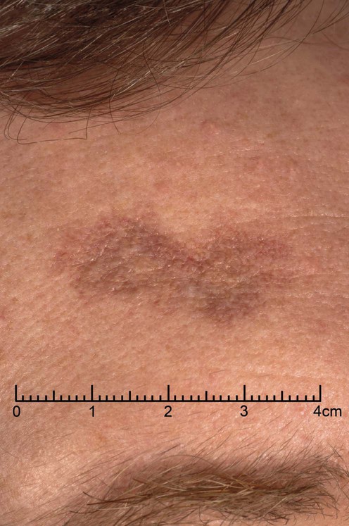
92 Granuloma faciale Nevianna Tomson and Susan E. Handfield-Jones Evidence Levels: A Double-blind study B Clinical trial ≥ 20 subjects C Clinical trial < 20 subjects D Series ≥ 5 subjects E Anecdotal case reports (© Addenbrooke’s Hospital.) Granuloma faciale is a rare, benign, chronic inflammatory dermatosis caused by a localized form of cutaneous vasculitis. It presents primarily in middle-aged Caucasian males, usually as a single lesion on the face. Multiple lesions occur in up to a third of patients and there are isolated reports of similar conditions affecting the eye and upper airways. Lesions are red-brown, violaceous or flesh-colored plaques or nodules with accentuation of follicular openings. Clinical diagnosis is difficult. Differential diagnosis includes sarcoid, lupus, lymphocytoma cutis, persistent insect bite reactions, and lymphoma. The histological differential diagnosis includes erythema elevatum diutinum, and angiolymphoid hyperplasia with eosinophilia. Management strategy Granuloma faciale is a chronic condition; spontaneous remission is unusual. Lesions are usually asymptomatic, but treatment is needed to reduce disfigurement. It is notoriously resistant to treatment and, because of the rarity of the condition, there are no formal trials of therapy. Treatment modalities can be divided into destructive techniques and anti-inflammatory approaches. The optimal treatment depends on the size, site, and thickness of the lesions. For isolated or small numbers of lesions intralesional steroid or destructive treatments such as cryotherapy, laser or surgical excision can be used. For multiple or widespread lesions systemic treatment, such as dapsone or clofazimine, can be considered. Cosmetic camouflage can be helpful for some patients with flatter lesions. Specific investigations Skin biopsy Hematology (complete blood count) Histological findings include a dense eosinophilic and neutrophilic infiltrate, often perivascular, affecting the upper and sometimes deep dermis. The epidermis is spared and there is a Grenz zone. Telangiectasia is common. Vasculitis with leukocytoclasis is reported. Dermal fibrosis is often seen. Granuloma faciale: a clinicopathologic study of 66 patients. Ortonne N, Wechsler J, Bagot M, Grosshans E, Cribier B. J Am Acad Dermatol 2005; 53: 1002–9. Peripheral blood eosinophilia is sometimes found. First-line therapies Corticosteroids E Cryotherapy D Laser therapy E Surgery E Only gold members can continue reading. Log In or Register to continue Related Related posts: Cat scratch disease Mucoceles Tinea capitis Herpes genitalis Necrolytic migratory erythema Nevoid basal cell carcinoma syndrome Stay updated, free articles. Join our Telegram channel Join Tags: Treatment of Skin Disease Comprehensive Therapeutic Strategies Aug 7, 2016 | Posted by admin in Dermatology | Comments Off on Granuloma faciale Full access? Get Clinical Tree
92 Granuloma faciale Nevianna Tomson and Susan E. Handfield-Jones Evidence Levels: A Double-blind study B Clinical trial ≥ 20 subjects C Clinical trial < 20 subjects D Series ≥ 5 subjects E Anecdotal case reports (© Addenbrooke’s Hospital.) Granuloma faciale is a rare, benign, chronic inflammatory dermatosis caused by a localized form of cutaneous vasculitis. It presents primarily in middle-aged Caucasian males, usually as a single lesion on the face. Multiple lesions occur in up to a third of patients and there are isolated reports of similar conditions affecting the eye and upper airways. Lesions are red-brown, violaceous or flesh-colored plaques or nodules with accentuation of follicular openings. Clinical diagnosis is difficult. Differential diagnosis includes sarcoid, lupus, lymphocytoma cutis, persistent insect bite reactions, and lymphoma. The histological differential diagnosis includes erythema elevatum diutinum, and angiolymphoid hyperplasia with eosinophilia. Management strategy Granuloma faciale is a chronic condition; spontaneous remission is unusual. Lesions are usually asymptomatic, but treatment is needed to reduce disfigurement. It is notoriously resistant to treatment and, because of the rarity of the condition, there are no formal trials of therapy. Treatment modalities can be divided into destructive techniques and anti-inflammatory approaches. The optimal treatment depends on the size, site, and thickness of the lesions. For isolated or small numbers of lesions intralesional steroid or destructive treatments such as cryotherapy, laser or surgical excision can be used. For multiple or widespread lesions systemic treatment, such as dapsone or clofazimine, can be considered. Cosmetic camouflage can be helpful for some patients with flatter lesions. Specific investigations Skin biopsy Hematology (complete blood count) Histological findings include a dense eosinophilic and neutrophilic infiltrate, often perivascular, affecting the upper and sometimes deep dermis. The epidermis is spared and there is a Grenz zone. Telangiectasia is common. Vasculitis with leukocytoclasis is reported. Dermal fibrosis is often seen. Granuloma faciale: a clinicopathologic study of 66 patients. Ortonne N, Wechsler J, Bagot M, Grosshans E, Cribier B. J Am Acad Dermatol 2005; 53: 1002–9. Peripheral blood eosinophilia is sometimes found. First-line therapies Corticosteroids E Cryotherapy D Laser therapy E Surgery E Only gold members can continue reading. Log In or Register to continue Related Related posts: Cat scratch disease Mucoceles Tinea capitis Herpes genitalis Necrolytic migratory erythema Nevoid basal cell carcinoma syndrome Stay updated, free articles. Join our Telegram channel Join Tags: Treatment of Skin Disease Comprehensive Therapeutic Strategies Aug 7, 2016 | Posted by admin in Dermatology | Comments Off on Granuloma faciale Full access? Get Clinical Tree


 Corticosteroids
 Cryotherapy
 Laser therapy
 Surgery


