(1)
Department of Plastic Surgery, Medical University of Gdańsk, Poland, Gdansk, Poland
Abstract
The use of free TRAM flaps for clinical and experimental purposes has a number of advantages. The rat rectus muscle has a predictable “flow-through” axial vascular system with six to seven perforators. Anatomy-wise, it is a reliable model with clearly defined limits and stable orientation points. It offers reliable blood supply, it is multifunctional, easy to prepare without subsequent complications. Its vessels (superior epigastric, in particular) have a diameter which is sufficient for an expert microsurgeon to perform anastomoses. It may be used in a number of laboratory studies on microcirculation, biochemical or pharmacological laboratory studies.
Keywords
RatRectus muscleFree tramAnimal modelMicrosurgeryComplicationsIntroduction
The last few decades have seen a significant development of techniques in reconstructive surgery, and micro-surgery in particular. It takes years of practice for novice surgeons to master the technical and manual skills and the process needs to be supervised by an experienced surgeon. Various models and simulators are available for practicing numerous diagnostic and therapeutic procedures. Training in laboratories with access to a range of experimental animal species, surgical tools and microscopes is a safe and effective method of developing the necessary expertise in prospective surgeons, who later use it in reconstructive procedures, including those involving microsurgical techniques. The choice of the experimental model is a critical factor in the success of a study [1–4].
Breast reconstruction following mastectomy is recognized as a step in the process of breast cancer treatment. Among the range of reconstructive treatment, autologous breast reconstruction is one of the methods which offer satisfactory results, both in terms of functional and aesthetic aspects [5, 6]. The basic method is a pedicled TRAM (Transverse Rectus Abdominis) flap [7]. Unfortunately, the flap, with its unreliable blood supply, often develops partial or total necrosis, resulting in the postponement of a possible adjuvant treatment and prolonged, often complicated wound healing. To prevent this type of complications most Reconstruction Institutions perform breast reconstruction with the free TRAM flap or its part without rectus abdominis muscle, which is then transmitted on the perforator vessels – DIEP flap type (Deep Inferior Epigastric Perforator) [8].
Flaps in Rat Models
Rats have been used as laboratory animals for a long time, making numerous medical research studies possible. It is a low-cost, easy-to-maintain animal, it is also relatively resistant to diseases [9, 10]. There are a number of rat varieties, which makes it possible to use the animal in a number of experimental projects offering an adequate chance of repeatability. In their work, Dunn and Mancoll [2] show the possibilities of applying a rat model in researching a number of tissue flaps. These include random pattern flaps from the tissues of the back, used in tissue necrosis studies, delay flaps used in research into delay phenomenon, also called McFarlane flap, named after their inventor [11]. Research conducted by other authors shows their disadvantages arising from dissimilar tissue vascularity in rats and humans in the back area [12, 13]. Frequently studies involve flaps from the pectoral and abdominal area which may have random and axial blood supply pattern derived from rectus abdominis muscles. Island flaps based on epigastric vessels may also be used in researching the impact of the microsurgical anastomoses on their function and survival. The first flap of this type was described in 1967 by Strauch and Murray, and a detailed description of the anatomy of the free lobe was presented as late as in 1984 by Petry and Wortham [14, 15], who used fluorescein to show the position of blood vessels and the survival of island flaps based on the inferior epigastric branch. These studies may be conducted on rats weighing at least 350 g, and the tissue flap should not exceed the area of 9 × 9 cm. Other free flap types which can be studied on a rat model include latissimus dorsi and external oblique flaps. These can be studied, however, on rats weighing at least 600 g. Composite flap research has gained particular significance in the recent years. They date back to the 1960s, when Buncke presented the model of a free tissue complex from a rat hindlimb without a part of femoral muscles [16]. Nowadays, transferring tissue complexes with the use of microsurgical anastomoses in rats has, in consequence, prepared the basis for subtotal transplants of the face and hand in people [3, 17].
Plastic and reconstructive surgery has greatly benefited from using laboratory rats in model studies analyzing the function and survival of tissue flaps. Thanks to a strong similarity in the anatomy of both species, it is a cheap and repeatable model, which can be translated onto human properties [18, 19].
Free TRAM Flap Model
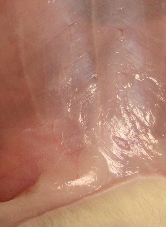
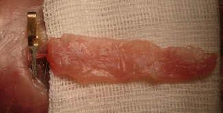
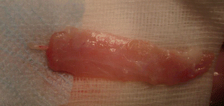
The value of the TRAM flap depends on its blood supply. Exploring the angiosomes concept [20, 21] and the development of microsurgical techniques have led to the discovery of new opportunities in tissue flap transfer. Free muscle flaps, myocutaneous and composite flaps (incorporating skin, muscle and bone tissue) have become the reality in reconstructive surgery. A number of those discoveries were possible thanks to experiments on animals [22, 23]. The animal model of microsurgical procedures contributes to research into flap survival, reinnervation, appropriate design and functioning. It is particularly significant because experiment results are, by and large, later reflected in the treatment of human patients. Due to the specific anatomical structure found in rats – panniculus carnosus – many of the available flaps do not always correspond to the human anatomy, which reduces their applicability in research. A rectus abdominis flap, however, shows a significant similarity in rats and humans, both in terms of two blood supplies – inferior and superior epigastric vessels, as seen in Figs. 7.1, 7.2, and 7.3, and the presence of perforator vessels providing blood supply for the skin, allowing for a broad experimental use [24].


Fig. 7.1
Inferior or caudal vascular pedicle of rat rectus muscle

Fig. 7.2
Superior or cranial vascular pedicle of rat rectus muscle

Fig. 7.3
Rat rectus free flap
The rectus muscle in rats extends on the entire surface of the abdomen, with its distal attachment at pubic symphysis, while the proximal one extends from the first rib, one-third medial of the clavicle, to the sternum. The blood supply in the rat muscle is similar to that in a human, has a double blood supply derived from inferior and superior epigastric vessels, and corresponds to type III vascular supply in Mathes and Nahai categories [25].
The first description of free TRAM flap use on a rat model comes from Zhang et al. and was presented in 1993 [26]. The study was divided into two stages. The first stage involved determining the anatomy of abdominal rectus muscle in a rat, and then the team performed a microsurgical anastomosis of superior epigastric vessels. In its stage focused on anatomy, the study showed the dominant role of blood supply from superior epigastric vessels with artery and vein diameter ranging from 0.4 to 0.5 mm, while the diameter of inferior arteries and veins is 0.2–0.3 mm, which, according to the authors, does not allow for the performance of anastomosis without common complications. The average length of a flap pedicle is 11 mm. The innervation of muscle is segmental and is derived from the nerves of ribs 5–13. Its disadvantage is the fact that nerve branches cannot be anastomosed during the transplantation procedure. The skin island of a free TRAM flap receives blood supply from six to seven myocutaneous perforating vessels. A study performed by Zhang [27


Stay updated, free articles. Join our Telegram channel

Full access? Get Clinical Tree


