and Peter M. Prendergast2
(1)
Elysium Aesthetics, Bogota, Colombia
(2)
Venus Medical, Dublin, Ireland
Liquid Gold
In the 1999 movie Fight Club, Tyler Durden, played by Brad Pitt, makes a living stealing fat from plastic surgery clinics and turning it into bars of soap. Who could foresee that the waste we throw away after every liposuction procedure could be recycled for another use?
Recycling industrial and human effluence has become an industry in itself. Fat is no exception. Some restaurants donate used animal and vegetable fat for use as biofuel for cars. Why not human fat? Although the prospect of using human adipose tissue as an energy source in industry seems bizarre, its use as an autotransplant in aesthetic surgery for beautification has been broadly accepted. To many surgeons, it has become liquid gold, allowing natural contouring of the body—even large surface areas—without many of the risks associated with silicone implants.
In 1893 Gustav Adolf Neuber first reported fat grafting as a surgical procedure at the 22nd meeting of the “Deutschen Gesellschaft für Chirurgie” in Berlin. The proceedings of this meeting were published in German only, and this limited the dissemination of fat grafting as an innovative technique to the non-German speaking world. In the 1980s, almost a century later, the idea of fat grafting gained new interest, particularly when liposuction techniques were popularized.
In the early 1980s, the Argentinian plastic surgeon Abel Chajchir conducted pioneering fat grafting procedures in humans [1, 2]. He designed instruments to aspirate fat, systems to freeze the fat, and techniques for subsequent grafting. This idea of volume replacement using one’s own fat was explored by only a few pioneers at the time, and generally disregarded or ignored by the rest of the medical community. Another visionary surgeon, Sydney Coleman, developed his technique of lipostructure and transformed fat grafting from an unreliable procedure to one that is safe, effective, and generally regarded as the gold standard in autologous fat transfer [3, 4]. Tiny pearls of fat are grafted using special blunt cannulae.
Zuk et al. reported the presence of stem cells in human adult fat in 2002 [5]. Since stem cells can differentiate into almost any type of cell, the promise of tissue regeneration from adipose-derived stem cells has become the subject of international research efforts. The fat that was once discarded after liposuction may be employed to restore damaged, aged, or atrophied tissues. Conditions such as diabetes, degenerative diseases, and even normal aging may be ameliorated by processing harvested lipoaspirate.
On the one hand, adipose tissue is regarded as undesirable and its excess is evident as the problem of obesity continues to grow worldwide. On the other hand, adipose tissue is acknowledged as essential for hormonal and metabolic processes and thermoregulation and for contributing to the beautiful curves and contours of the human form. International organizations, such as the International Federation for Adipose Therapeutics and Science, run dedicated educational and scientific activities devoted to fat.
Fat Metabolism and Endocrinology
Fatty tissue is the largest energy reserve in the body. At birth, fat represents about 17 % of bodyweight; in adolescence, around 20 % in women; and 10 % in men with some phenotypic variations. In normal adults, fat approaches 15–20 % in men and 25–30 % in women. Two types of fat are described in the human body: white adipose tissue and brown adipose tissue.
The distribution of body fat varies between populations in different countries. However, fat excess in the upper body is often associated with certain disease states, especially metabolic disorders. Fat distribution is determined by a complex interplay between central regulation in the hypothalamus, receptors in the adipose tissue, and the various molecules that interact with these receptors. Catecholamines, insulin, growth hormone, steroids, and diverse neuropeptides modulate activity at these receptors. Lipogenesis and lipolysis are regulated by lipase lipoprotein and hormone-sensitive lipase, respectively. This system is regulated mainly in the hypothalamus where an assemblage of regulatory centers governs homeostasis. The ventromedial nucleus of the hypothalamus serves as a satiety center and the lateral hypothalamic area serves as a feeding center. They coordinate the processes that govern eating behavior and the subjective perception of satiety. Further, the secretion of hormones from the thyroid gland, adrenal gland, and pancreatic islet cells is influenced by these areas in response to metabolic stress.
The hypothalamus regulates caloric intake, utilization, and storage in a manner that tends to maintain the body weight in adulthood. Although the biochemical mechanisms that control fat deposits are well recognized, the presumptive set point around which it attempts to stabilize body weight is poorly defined or maintained, as it changes readily with changes in physical activity, composition of the diet, emotional states, stress, pregnancy, and so on. Carbohydrate metabolism is functionally related to lipogenesis in the adipose tissue. Both processes are regulated in the liver in order to obtain the energy from glucose the body needs in different metabolic situations.
Mechanisms involved in obesity-related pathologies include increased fatty acid input to the portal circulation, tumor necrosis factor and angiotensin II production, and steroidogenesis. Adipocyte insulin resistance may be implicated in the complex pathogenesis of obesity.
White fat cells (adipocytes) produce leptin, a key hormone in the regulation of body weight. Starvation corresponds to low leptin levels, while obesity corresponds to high levels of leptin. The hypothalamus specifically controls the feeding process depending on the signals received from the cells in the gastrointestinal tract and on the leptin levels.
Primarily, brown fat cells play a thermoregulatory role in mammals. However, thermogenic defects observed in some obesity models in animals do establish a link with the development of obesity. In humans, brown fat cells play an important role in heat production, at least in the perinatal period. However, the complete role of brown fat is not fully understood. The possibility to pharmacologically stimulate these fat cells makes them a potential target for therapeutic intervention in obesity.
Fatty tissue also plays an important role as an energy source during exercise. Fat, the major energy store in the body, is mobilized from adipose tissue as free fatty acids to provide metabolic fuel. At lower exercise intensities, fats may provide 50–60 % of the energy for muscle contraction, but this process cannot keep pace with the high demands for energy that occur during heavy exercise. Energy liberation from fat is slower than the liberation of glucose from glycogen. Moderate activity, in fact, favors the consumption of fat as muscle fuel. The depletion of body fat reserves is almost never a limiting factor in muscle activity. In the absence of other fuels, proteins can serve as an energy source for contraction, particularly as it occurs during heavy, prolonged, or intense exercise.
Fat Anatomy: The Subcutaneous Tissue and Superficialis Fascia
The subcutaneous tissue is an organ that has received limited attention by anatomists. Is has been widely known as a three-layer system divided by a membranous tissue. This tissue layer has received many names such as superficial fascia, Scarpa’s fascia, Colles’ fascia, and Camper’s fascia [6–9]. The varied nomenclature can be somewhat confusing.
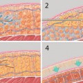
Current classification divides this structure into three layers: a superficial adipose tissue layer (SAT), an intermediate membranous layer (superficialis fascia), and a deep adipose tissue layer (DAT) (Fig. 7.1) [8, 9].

Fig. 7.1
Fat anatomy. Composed of two main layers: superficial and deep. A third intermediate layer is composed of the superficial fascia
The SAT layer is characterized by fibrous septa that define polygonal lobes of fat tissue. The thickness of this layer is relatively constant throughout the body. It is this layer that correlates with the pinch test that is commonly used to gauge the depth of insertion of the cannula for suction lipectomy.
The intermediate membranous layer is a continuous fibrous membrane rich in elastic fibers.
Related posts:
Stay updated, free articles. Join our Telegram channel

Full access? Get Clinical Tree


