Fig. 4.1
Female rat genitourinary system
Technique
The uterus is approached through a midline incision extending from the xiphoid process to the pubic symphysis with care taken to avoid the bladder. Two retractors are used to open the surgical site and to allow good visualization. The intestines can be placed on a gauze and retracted to one side. The gauze should be occasionally moistened with a 0.9 % saline solution. The base of the uterus lies just posteriorly to the bladder. If the bladder is full, it may be emptied by direct needle aspiration to improve exposure and to prevent urine dribbling during the surgery. Two uterine horns should be easily visible.
The horn is transected sharply with iris scissors at the middle third of its length. In clinical practice, the occluded tubal segment is usually excised from the adjacent structures, thus creating a defect in the mesosalpinx. In the training rat model, it may be desirable to transect a small portion of the mesosalpinx to most closely simulate a clinical scenario. Hemostasis is achieved by prompt coagulation of the more significant bleeders with micro-bipolar forceps or ophthalmic cautery. Bleeding is usually located at the mesosalpinx and between the serosa and muscularis. Overzealous coagulation of the mesosalpinx and horn epithelium should be avoided to prevent devitalizing the anastomosis site, which will adversely affect its function. Frequent irrigation is performed with Lactated Ringer’s or normal saline with 50 units of heparin per 10 ml of solution. Before anastomosis, the cut surface should be thoroughly examined under high magnification to ensure normal anatomical architecture with intact mucosal folds.
When there is no significant luminal disparity, the distal and proximal portions of the uterus are prepared for anastomosis. Both segments are approximated in two layers. The first layer joins the epithelium and the muscularis and the second one joins the serosa. The sutures used in fallopian tube repair are usually 7-0 or 8-0 polyglactin, polypropylene or nylon on a 100–150 μm needle. There is still controversy over the ideal suture material for microsurgical tube anastomosis. In clinical practice both absorbable and nonabsorbable sutures have been used with excellent results. Several experimental studies conducted on rabbit fallopian tubes and rat uterine horns showed that the type of suture material did not influence the pregnancy rate or patency of the anastomosis. However, the use of nonabsorbable sutures significantly altered the histologic structure and produced greater tissue reaction [5, 6, 17, 18].
The repair is started with a single suture placed in the mesosalpinx close to the tube to align both uterine segments and to reduce tension from the anastomosis. Then the inner layer of anastomosis, joining epithelium and muscularis, is completed with interrupted sutures as described by Gomel [2]. All knots should be positioned extraluminally. The first musculoepithelial suture is placed at the mesosalpingeal border at 6-o’clock position to ensure proper alignment of both uterine segments (Fig. 4.2). When this suture is tied, two or more additional equally spaced stitches (depending on the type of anastomosis) are placed to appose the inner layer. The sutures should be tied loosely to prevent strangulation. It may also be preferable to tie the knots after all sutures have been placed, particularly when visibility of the lumen is limited (Fig. 4.3). Whether or not the lumen should be included remains controversial and currently there is no sufficient data to favor any of the two options [19].
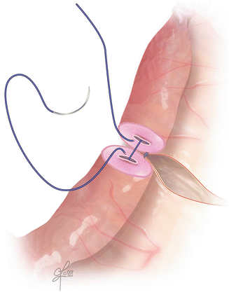
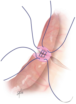
Fig. 4.2
The first musculoepithelial suture placed at the mesosalpingeal border at 6-o’clock position to ensure proper alignment of both uterine segments
Fig. 4.3
Three or four stitches are placed to complete the inner layer of the anastomosis. It may be preferred to tie the knots after all sutures have been placed, particularly when visibility of the lumen is limited
After approximation of the first layer the patency test should be performed. A dye solution is injected into the uterine horn and its spill is observed at the other end. The anastomosis does not have to be watertight as long as spill of dye is seen distally. Additional stitches may be needed if there is a leak at the anastomosis site but no distal spill. The serosa can be joined with four to eight equally spaced interrupted stitches (Fig. 4.4). Alternatively, two continuous sutures, starting at the antimesosalpingeal boarder at 12-o’clock and running on the anterior and posterior walls, may be used. Finally, the defect in the mesosalpinx is repaired with simple interrupted sutures (Fig. 4.5).

 Microcirculation Model for Invasive Animal Monitoring
 Epineural Sleeve End-to-End Repair
 Somatosensory Evoked Potential Model for Assessment of Nerve Regeneration
 Peripheral Nerve Surgery Models Crush Injury and Epineural Patch
 Heterotopic Vascularized Ovarian Autotransplantation Model in the Sheep
 Defect Repairs After Resections of Laryngeal Cancer, Hypopharyngeal Cancer, and Cervical Esophageal Cancer


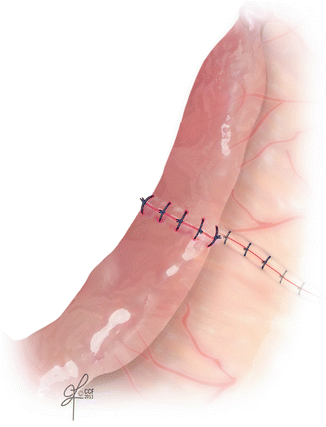
Fig. 4.4
The serosa is joined with four to eight equally spaced interrupted stitches. Alternatively, two continuous sutures, starting at the antimesosalpingeal boarder at 12-o’clock and running on the anterior and posterior wall may be used
Related posts:
Microcirculation Model for Invasive Animal Monitoring
Epineural Sleeve End-to-End Repair
Somatosensory Evoked Potential Model for Assessment of Nerve Regeneration
Peripheral Nerve Surgery Models Crush Injury and Epineural Patch
Heterotopic Vascularized Ovarian Autotransplantation Model in the Sheep
Defect Repairs After Resections of Laryngeal Cancer, Hypopharyngeal Cancer, and Cervical Esophageal Cancer
Stay updated, free articles. Join our Telegram channel
Full access? Get Clinical Tree
