Excision of Soft Tissue Tumors of the Knee
Raffi S. Avedian
DEFINITION
Soft tissue sarcomas are malignant tumors that arise from mesenchymal cells and are classified according to their cell of origin such as muscle, tendon, or fat.
Peripheral nerve sheath tumors arise from neural crest cells but are typically grouped with sarcomas because of similarities in location, natural history, and treatment.
The World Health Organization groups soft tissue sarcomas into 10 categories based on the tissue of origin.
Most sarcomas occur in a deep location, meaning below the muscle and fascia, whereas one-third of sarcomas occur in a subcutaneous location.
Patients with soft tissue sarcomas and patients with benign tumors often present with the same chief complaint of having a soft tissue mass.
Rendering an accurate diagnosis prior to surgery is important because the treatment plan may vary based on the nature of the diagnosis. For example, a benign tumor may be monitored or removed with a simple excision, whereas in the case of a sarcoma, the goal of surgery is to remove the tumor with a wide margin. With appropriate planning, radiotherapy and/or chemotherapy is often incorporated into the treatment plan either before or after definitive surgery.
ANATOMY
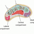
The knee is the largest joint in the body. It is made up of the patellofemoral, lateral, and medial compartments.
Knee motion is primarily flexion and extension; however, bending and rotation are important components of knee kinematics.
The primary stabilizers of the knee are the extra-articular medial and lateral cruciate ligaments and intra-articular anterior and posterior cruciate ligaments.
The knee has several layers of covering including the capsule and retinacular tissues. During tumor resection, superficial layers may be used as the deep margin for the tumor, whereas the deeper capsule may be preserved to keep the joint closed.1
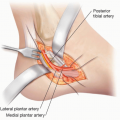
The popliteal artery travels along the back of the knee and along with the tibial nerve travels between the lateral and medial head of the gastrocnemius. It branches into the posterior tibial artery, which travels deep to the soleus muscles; the anterior tibial artery, which passes from posterior to the anterior compartment distal to the tibia-fibula joint; and the peroneal artery, which branches off the tibiofibular trunk and is located medial to the fibula next to the flexor hallucis longus.
The geniculate vessels branch off from the popliteal vessels and are often all ligated during tumor resection of the distal femur.
The tibial nerve is located next to the popliteal vessels. The common peroneal nerve travels medial and deep to the biceps femoris muscle, a constant relationship that facilitates finding and protecting the nerve during tumor dissection.
The common peroneal nerve wraps around the neck of the fibula and divides into the deep branch that innervates the anterior muscle compartment and the superficial branch that innervates the lateral compartment.
The lateral sural cutaneous nerve, which branches off the common peroneal nerve, may be large in some patients and be confused with the common peroneal nerve.
PATHOGENESIS
The mechanism for sarcoma formation is not known.
Risk factors for sarcoma development include radiation exposure such as medical radiotherapy, pesticide exposure, and hereditary conditions including Li-Fraumeni syndrome and neurofibromatosis.
NATURAL HISTORY
All sarcomas have the potential for local recurrence and metastasis.
Tumor variables that are associated with increased risk of metastasis include high grade and large size (greater than 5 cm).
Lungs are the most common location of metastasis.
PATIENT HISTORY AND PHYSICAL FINDINGS
Determining when the mass was first noticed and how rapidly it is growing can help the clinician differentiate between benign and malignant tumors.
The presence of pain is often associated with a benign tumor, such as a schwannoma or vascular malformation, and may not be present with sarcomas until late in the disease course.2
Determining the size of the tumor, manual muscle strength testing, and sensory examination are useful to determine if there is a neurological compromise. Limb edema assessment and pulse examination can help determine if the tumor is causing vascular or lymphatic compromise.
Range of motion testing and gait assessment are helpful in assessing a patient’s mobility and functional status to guide perioperative and postoperative counseling.
IMAGING
Plain radiographs can help diagnose joint degeneration or jointbased lesions such as gout and synovial osteochondromatosis.
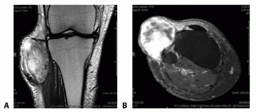
FIG 1 • A. Coronal and axial (B) MRIs demonstrating a high-grade soft tissue sarcoma abutting the proximal tibia and knee joint.
Magnetic resonance imaging is the modality of choice for soft tissue tumor evaluation (FIG 1A,B).Related posts:
 Fasciotomy of the Thigh, Lower Leg, and Foot
 Bony Reconstruction of Foot and Ankle (Bone Grafts)
 Vascular Reconstruction of Lower Extremity, Foot, and Ankle
 Tibial Reconstruction
 Reconstruction of Femur
 Amputation of the Lower Extremity: Above-Knee Amputation, Below-Knee Amputation, Through-Knee Amputation

Stay updated, free articles. Join our Telegram channel

Full access? Get Clinical Tree


