Abstract
Dermatology is best approached by classifying cutaneous and oral disorders by the primary lesion (e.g., macule, tumor) and the distribution of lesions (e.g., facial, axillary, and oral). Primary lesions are the initial lesion that has not been altered by trauma, secondary lesions (e.g., excoriation), or natural regression. This introductory chapter organizes mucocutaneous disorders by their primary lesion and distribution to aid in the development of broad differential diagnosis.
Key words
Macule, papule, plaque, nodule, alopecia, pustule, tumor, vesicle, bulla, erosion, ulcer, desquamation
A
History and Physical Examination
- •
The initial step in the dermatologic evaluation involves obtaining a detailed dermatologic history. Box 1.1 describes pertinent questions.
BOX 1.1
Dermatologic History
From Goldstein BG, Goldstein AO: Practical Dermatology, ed 2, St. Louis, 1997, Mosby.
A
Initial Questions
- 1.
When did the rash start?
- 2.
What did it look like when it first started, and how has it changed?
- 3.
Where did it start, and where is it located now?
- 4.
What treatments, especially over-the-counter medications or self-remedies, has the patient tried? What was the effect of each of these treatments?
- 5.
Are there symptoms (e.g., itching, pain)?
- 6.
What is the patient’s main concern about the rash (e.g., itching, pain, cancer)?
- 7.
How is the rash affecting the patient’s life?
- 8.
Are other family members concerned or affected?
- 9.
Has the patient ever had this rash before? If so, what treatment worked?
- 10.
What does the patient think caused the rash?
B
Follow-up Questions
- 1.
Does the patient have a history of chronic medical problems?
- 2.
What is the patient’s social history, including occupation (chemical exposures), hobbies, alcohol and tobacco use, and any underlying interpersonal or family stress?
- 3.
What medications is the patient taking, acutely or chronically, including birth control pills and over-the-counter medications?
- 4.
Does the patient have any underlying allergies?
- 5.
Is there a family history of hereditary or similar skin diseases?
- 6.
Will the patient’s education or financial status influence treatment considerations?
- 1.
- •
When examining the patient, it is essential to accurately and concisely describe the skin lesions, their distribution, and their overall characteristics.
- •
Skin lesions should be classified as primary or secondary:
- •
Primary lesions represent the initial lesional morphology, and are critical to recognize in order to reach an accurate diagnosis.
- •
Secondary lesions may result from evolution or chronicity of the primary lesion, or may be created by scratching, infection, and other secondary skin changes.
- •
- •
The proper terminology in describing these lesions is described in Boxes 1.2 and 1.3 .
BOX 1.2
Primary Skin Lesions
From Goldstein BG, Goldstein AO: Practical dermatology , ed 2, St. Louis, MO, 1997, Mosby.
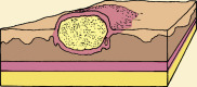
Macule : Small spot, different in color from surrounding skin, that is neither elevated nor depressed below the skin’s surface
Papule: Small (≤5 mm diameter) circumscribed solid elevation of skin
Plaque: Large (≥5 mm) superficial flat lesion, often formed by a confluence of papules
Nodule: Large (5–20 mm) circumscribed solid skin elevation
Pustule: Small circumscribed skin elevation containing purulent material
Vesicle: Small (<5 mm) circumscribed skin blister containing serum
Wheal: Irregular elevated edematous skin area, which often changes in size and shape
Bulla: Large (>5 mm) vesicle containing free fluid
Cyst: Enclosed cavity with a membranous lining, which contains liquid or semisolid matter
Tumor: Large nodule, which may be neoplastic
Telangiectasia: Dilated superficial blood vessel
BOX 1.3
Secondary Skin Lesions
From Goldstein BG, Goldstein AO: Practical dermatology , ed 2, St. Louis, MO, 1997, Mosby.
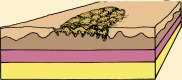
Scale: Superficial epidermal cells that are dead and cast off from the skin
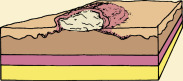
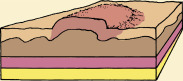
Erosion: Superficial, focal loss of part of the epidermis; lesions usually heal without scarring
Ulcer: Focal loss of the epidermis extending into the dermis; lesions may heal with scarring
Fissure: Deep skin split extending into the dermis
Crust: Dried exudate, a “scab”
Erythema: Skin redness
Excoriation: Superficial, often linear skin erosion caused by scratching
Atrophy: Decreased skin thickness due to skin thinning
Scar: Abnormal fibrous tissue that replaces normal tissue after skin injury
Edema: Swelling due to accumulation of water in tissue
Hyperpigmentation: Increased skin pigment
Hypopigmentation: Decreased skin pigment
Depigmentation: Total loss of skin pigment
Lichenification: Increased skin markings and thickening with induration secondary to chronic inflammation caused by scratching or other irritation
Hyperkeratosis: Abnormal skin thickening of the superficial layer of the epidermis
- •
For diagnostic purposes, it is also important to note the distribution of the skin lesions, as many dermatologic conditions present in characteristic anatomic locations or in specific configurations.
- •
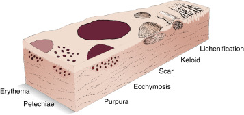
Table 1.1 describes vascular and miscellaneous skin dermatoses.
TABLE 1.1
Vascular Skin Lesions
From Swartz MH: Textbook of physical diagnosis: history and examination , ed 6, Philadelphia, 2010, Saunders.
Lesion
Characteristics
Examples
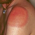
Erythema
Pink or red blanchable discoloration of the skin secondary to dilatation of blood vessels
Facial flushing
Petechiae
Reddish-purple; nonblanching; smaller than 0.5 cm
Intravascular defects
Purpura
Reddish-purple; nonblanching; greater than 0.5 cm
Intravascular defects
Ecchymosis
Reddish-purple; nonblanching; variable size
Trauma, vasculitis
Telangiectasia
Fine, irregular dilated blood vessels
Dilatation of capillaries
Spider Angioma
Central red body with radiating spider-like arms that blanch with pressure to the central area
Liver disease, estrogens
Miscellaneous Skin Lesions
Lesion
Characteristics
Examples
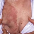
Scar
Replacement of destroyed dermis by fibrous tissue; may be atrophic or hyperplastic
Healed wound
Keloid
Elevated, enlarging scar growing beyond boundaries of wound
Burn scars
Lichenification
Roughening and thickening of epidermis; accentuated skin markings
Atopic dermatitis
B
Dermatoses by Anatomic Region
1
Scalp
Papules/Plaques
- •
Actinic keratosis
- •
Appendageal tumor
- •
Cyst
- •
Hemangioma
- •
Lichen planopilaris
- •
Lupus erythematosus
- •
Melanoma
- •
Nevus
- •
Seborrheic keratosis
Nodules
- •
Actinic keratosis
- •
Appendageal tumor
- •
Basal cell carcinoma
- •
Cyst
- •
Hemangioma
- •
Kerion
- •
Metastatic carcinoma
- •
Nevus
- •
Prurigo nodularis
- •
Seborrheic keratosis
Eruptions
- •
Contact dermatitis
- •
Dissecting cellulitis
- •
Eczema
- •
Folliculitis
- •
Herpes zoster
- •
Pediculosis capitis
- •
Psoriasis
- •
Seborrheic dermatitis
- •
Tinea capitis
Alopecias
- •
Alopecia areata
- •
Anagen effluvium
- •
Androgenetic alopecia
- •
Discoid lupus erythematosus
- •
Hypervitaminosis A
- •
Lichen planopilaris
- •
Syphilis
- •
Systemic disease
- •
Telogen effluvium
- •
Tinea capitis
- •
Traction/chemical alopecia
- •
Trichotillomania
2
Face
Isolated Papules
- •
Acrochordon
- •
Actinic keratosis
- •
Angioma
- •
Appendageal tumors
- •
Basal cell carcinoma
- •
Cyst
- •
Dermatosis papulosa nigra
- •
Hemangioma
- •
Keratoacanthoma
- •
Lentigo maligna
- •
Milia
- •
Nevus
- •
Sebaceous hyperplasia
- •
Seborrheic keratosis
- •
Solar lentigo
- •
Squamous cell carcinoma
- •
Telangiectasia
- •
Venous lake
- •
Xanthelasma
Eruptions
- •
Acne rosacea
- •
Acne vulgaris
- •
Angiofibroma (Adenoma sebaceum)
- •
Dermatomyositis
- •
Eczema, including contact dermatitis
- •
Erysipelas
- •
Favre-Racouchot (comedones in actinically damaged skin)
- •
Fifth disease
- •
Herpes simplex/zoster
- •
Impetigo
- •
Lupus erythematosus
- •
Lymphocytoma cutis
- •
Melasma
- •
Pemphigoid/pemphigus
- •
Perioral dermatitis
- •
Photodrug eruption
- •
Pityriasis alba
- •
Postinflammatory hypopigmentation
- •
Psoriasis
- •
Sarcoidosis
- •
Scleroderma
- •
Seborrheic dermatitis
- •
Steroid rosacea
- •
Syphilis
- •
Tinea corporis
- •
Urticaria, angioedema
- •
Warts, especially flat or molluscum
3
Oral Mucosa
Oral Mucosa (See also “Erosions and Ulcers”)
- •
Kaposi’s sarcoma
- •
Leukoplakia
- •
Melanoma
- •
Mucous cysts
- •
Oral hairy leukoplakia
- •
Oral melanotic macule
- •
Pyogenic granuloma
- •
Verruca
4
Axilla
- •
Acanthosis nigricans
- •
Acrochordon
- •
Axillary freckling in neurofibromatosis
- •
Bullous pemphigoid
- •
Contact dermatitis
- •
Epidermal inclusion cyst
- •
Erythrasma
- •
Fox-Fordyce disease
- •
Fungal or yeast infection
- •
Hailey-Hailey disease
- •
Hidradenitis suppurativa
- •
Intertrigo
- •
Pediculosis corporis
- •
Pseudoxanthoma elasticum
- •
Scabies
- •
Striae distensae
- •
Trichomycosis axillaris
5
Hands and Feet
Isolated Papules
- •
Actinic keratosis
- •
Arsenical keratosis
- •
Basal cell carcinoma
- •
Callus/clavus
- •
Felon
- •
Keratoacanthoma
- •
Melanoma
- •
Nevus
- •
Painful fat herniations
- •
Pyogenic granuloma
- •
Solar lentigo
- •
Squamous cell carcinoma
- •
Warts
Eruptions
- •
Acute or chronic paronychia
- •
Cutaneous larva migrans (feet)
- •
Dermatomyositis
- •
Drug eruption
- •
Eczema, including contact dermatitis
- •
Emboli
- •
Epidermolysis bullosa
- •
Erythema multiforme
- •
Granuloma annulare
- •
Hand-foot-and-mouth disease
- •
Herpetic whitlow
- •
Hyperhidrosis
- •
Juvenile plantar dermatosis
- •
Keratolysis exfoliativa
- •
Lichen planus (wrists, ankles)
- •
Lupus erythematosus
- •
Pitted keratolysis (feet)
- •
Pityriasis rubra pilaris
- •
Porphyria cutanea tarda
- •
Psoriasis
- •
Reiter’s syndrome
- •
Rocky Mountain spotted fever
- •
Scabies
- •
Scleroderma
- •
Syphilis
- •
Tinea pedis, manus
- •
Viral exanthems
- •
Vitiligo
6
Genitalia/Inguinal
- •
Acrochrodons
- •
Accrodermatitis enteropathica
- •
Angiokeratoma
- •
Balanitis
- •
Bowen’s disease
- •
Candidiasis
- •
Chancroid
- •
Condyloma accuminata
- •
Contact dermatitis
- •
Diaper dermatitis
- •
Erythema multiforme
- •
Erythrasma
- •
Fixed drug eruption
- •
Folliculitits
- •
Furunculosis
- •
Herpes simplex/zoster
- •
Hidradenitis suppurativa
- •
Intertrigo
- •
Kawasaki syndrome
- •
Lichen planus
- •
Lichen sclerosus
- •
Lichen simplex chronicus
- •
Lymphogranuloma venereum
- •
Molluscum contagiosum
- •
Paget’s disease, extramammary
- •
Pearly penile papules
- •
Pediculosis pubis
- •
Perianal streptococcal cellulitis
- •
Pinworm
- •
Pityriasis rubra pilaris
- •
Psoriasis
- •
Reiter’s syndrome (reactive arthritis)
- •
Scabies
- •
Seborrheic dermatitis
- •
Squamous cell carcinoma
- •
Syphilis
- •
Tinea cruris
7
Photodistributed
- •
Dermatomyositis
- •
Lupus erythematosus
- •
Pellagra
- •
Photodrug eruption
- •
Polymorphous light eruption
- •
Porphyria cutanea tarda
Related posts:

Stay updated, free articles. Join our Telegram channel

Full access? Get Clinical Tree


