Summary and Key Features
- •
Cosmeceutical formulations are tested through noninvasive techniques to ensure consumer efficacy.
- •
2-Dimensional and 3-dimensional photography and image analysis are used to analyze surface texture, wrinkles, and aspects of skin tone such as evenness of tone and hyperpigmentation.
- •
Transepidermal water loss is a measure of the water leaving the skin to the atmosphere and can be quantified via a combination of precise temperature and humidity sensors. This is a direct measurement of the skin’s barrier function and the ability of cosmeceuticals to impact or supplement the skin barrier, either positively or negatively.
- •
Measuring the electrical properties of the skin, such as its capacitance or conductance, is useful in assessing the skin’s hydration and the moisturizing properties of cosmeceuticals.
- •
Colorimeters are used to measure various aspects of skin color such as melanin and erythema.
- •
Biomechanical properties such as elasticity and firmness are measured via various instruments which push, pull, and twist the skin, quantifying the stresses applied and strains produced.
- •
Diffuse reflectance spectroscopy (DRS) and laser Doppler velocimetry are used to assess cutaneous blood flow changes with DRS being able to measure changes in melanin as well.
- •
High-frequency ultrasound is used to gain insight into skin structure, such as the thickness of the skin and the density of connective tissue, without invasive biopsy procedures.
- •
Other optical techniques using high-power white light or low-power laser light, such as optical coherence tomography, Raman spectroscopy, and fluorometer are used to evaluate the molecular composition of the skin and the penetration of cosmeceutical ingredients into the skin.
See .
Introduction
This chapter is intended to provide an introductory survey of instrumental methods for evaluating cosmeceutical efficacy on human skin. Although the emphasis will be on instrumental methods, it is strongly recommended that a three-pronged approach that includes the instrumental methods along with clinical evaluations and panelists’ self-assessments is utilized whenever possible to evaluate the effects of various cosmeceuticals on skin condition.
We will begin by discussing those methods that measure aspects of the skin that are directly related to how the dermatologist and/or patient evaluate skin condition—that is, primarily to look with the eyes and feel with the fingers. Other instrumental techniques measure properties that cannot readily be appreciated by either visual or tactile means. These include assessments based on physiologic processes such as blood flow and transepidermal water loss rates.
Visual, Optical, and Laser-Based Methods
Imaging and Image Analysis
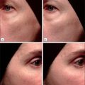
One of the more popular categories of claims currently being made for many cosmeceuticals is antiaging claims, such as “reduces the appearance of fine lines and wrinkles” or “reduces uneven skin tone.” Although such changes can be documented by standardized clinical photographs, it is desirable to quantify them more precisely than is achievable via clinical evaluation and self-assessment. A historically popular method of measuring changes in wrinkles, having been used for decades to successfully evaluate product effects, is to cast a replica of the skin surface using a silicone rubber impression material and to analyze the replica using a technique known as optical profilometry. This method can reliably quantify aspects of skin texture and wrinkle depth, but it is messy and difficult to master. With improvements in clinical photography, such as higher-resolution images, well-controlled lighting, faster image acquisition times, and three-dimensional imaging, better options have emerged.
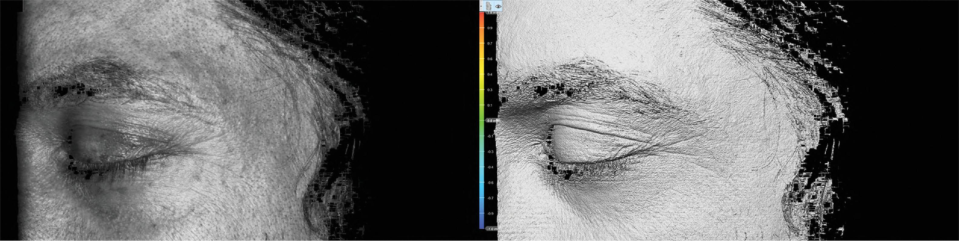
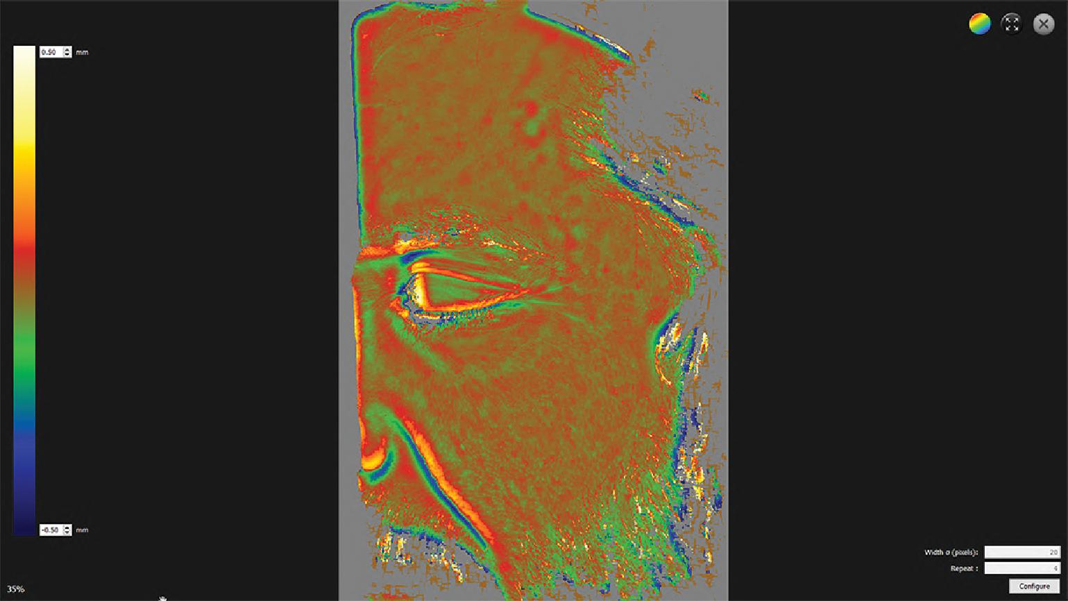
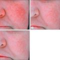
Objective data related to wrinkle length and depth can be calculated from two-dimensional images by measuring the area of shadows cast by high-raking light, or light shining on the subject at an angle from above. Three-dimensional imaging can also be used to capture data on texture and wrinkle depth. There are myriad methods of acquiring three-dimensional images, the most common being fringe projection. Fringe projection involves capturing two sets of images from fixed positions simultaneously while a projector displays patterns onto the skin. The associated software then calculates the distortion of the patterns and how they differ between the images from the two cameras to generate images with X, Y resolution as high as 20 µm and Z resolution as high as 2 µm ( Figs. 3.1 and 3.2 ). These systems tend to capture images more quickly, as they are not reliant on a flash, and they are less sensitive to changes in lighting, as analysis is not based on lighting intensity but rather on distortion of the visible patterns projected onto the surface. They are also less sensitive to changes in positioning, as landmarks can be used in the images during analysis to align them in three dimensions.
More recently dynamic facial imaging systems have been developed that capture video of a subject while they move their face between a resting position and an expression such as a grimace or a smile. This technique captures more information related to expression lines that occur through repetitive or sustained stress on the skin from the facial muscles and may be treated separately from texture, such as through Botox injection.
Texture analysis is but one example of how computerized image analysis can be used to objectively extract quantitative information from images. Box 3.1 provides a listing of some of the more common applications of image analysis that have been used to study skin structure and function. The basic rule applies that anything that can be seen by the unaided eye can easily be measured. Moreover, by using specialized lighting techniques such as polarization and illumination with ultraviolet (UV) or fluorescent light, things that cannot be directly visualized can be detected and measured via imaging.
- •
Wrinkle and texture analysis
- •
Changes in pigmentation and color
- •
Changes in reflectance (e.g., radiance, dullness, shine)
- •
Psoriasis lesions
- •
Acne lesions
- •
Weal and flare response
- •
Wounds and ulcers
- •
Sticky tape specimens/D-Squame discs
- •
Exfoliative cytology
- •
Sebutape specimens
- •
Sweat gland patterns
Skin Coloration
Another important visual clue to the condition of the skin after cosmeceutical application is its color, which depends on a number of factors including pigmentation, blood perfusion, and desquamation patterns. Experienced dermatologists frequently use color information in several ways. First, they can certainly appreciate the distribution of erythema and/or pigmented lesions on the basis of color. Moreover, by evaluating changes in the hue and/or intensity of color over time, they can tell whether patients are responding to treatment. Although the human eye is very sensitive, especially in detecting very subtle differences in contrast, the evaluation of color is still highly subjective. Color-measuring devices offer the advantages of objectivity and quantification on a continuous scale that can be referenced to color standards.
The devices that are currently being employed in experimental dermatology, skin pharmacology, toxicology, and cosmetic science to measure skin color changes fall into two distinct instrument types, as shown in Box 3.2 . In one category we have the tristimulus colorimeters, which are based on the three-dimensional L*a*b* color space (CIELAB). L*a*b* allows any color to be mathematically described by its hue (position on the color wheel), value (lightness), and chroma (saturation). Examples of these devices include the Chroma Meter (Minolta) and the Dr. Lange Micro Color (Dr. Bruno Lange GmbH), which have seen widespread use for quantifying erythema in the study of irritant dermatitis due to exposure to detergents, determining topical corticoid activity in the vasoconstriction test, and measuring the percutaneous penetration of vasodilators such as nicotinic acid.
Related posts:


Stay updated, free articles. Join our Telegram channel

Full access? Get Clinical Tree


