Histopathology:
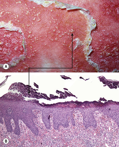
Abundant neutrophils (arrow) below the stratum corneum (Fig. 7.3); may be indistinguishable from acute generalized exanthematous pustulosis and subcorneal pustular dermatosis
Pustular Psoriasis, Variants
Localized – pustules limited to plaques of psoriasis (Fig. 7.4)
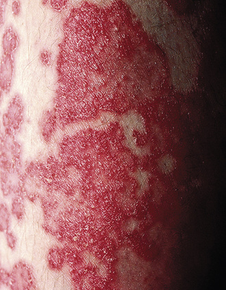
Annular – rings of erythema studded with peripheral pustules (Fig. 7.5)
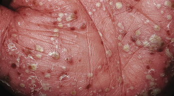
Palmoplantar (Fig. 7.6)
Acrodermatitis continua of Hallopeau – distal digit with erythema/scale/pustules (see Fig. 4.11B)
Acute Generalized Exanthematous Pustulosis
Commonly induced by antibiotics (penicillins, macrolides)
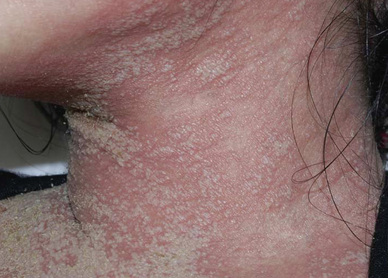
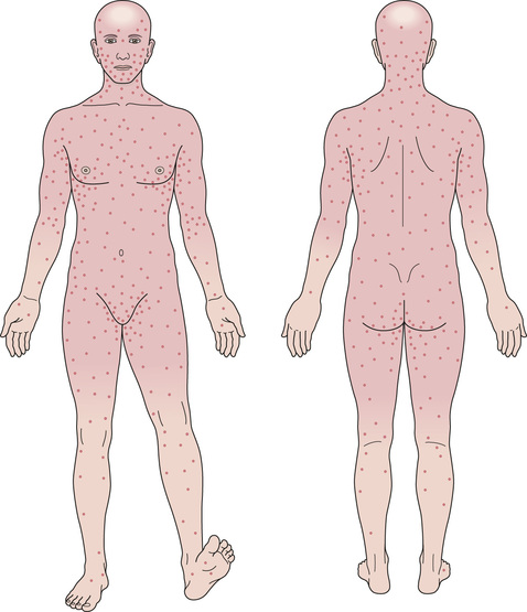
Begins on the face/body folds and becomes generalized (Fig. 7.7)
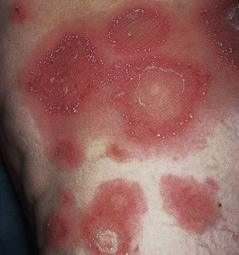
Small, sterile pustules over edema and erythema (Figs 7.8, 7.9)
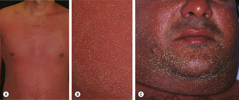
Fig. 7.8 Acute generalized exanthematous pustulosis. C, Courtesy, Kalman Watsky, MD. A,B, From Min JA, Park HJ, Cho BK, Lee JY. Acute generalized exanthematous pustulosis induced by Rhus (lacquer). J Am Acad Dermatol. 2010;63:166–8, © Elsevier. C, From Bolognia JL, Jorizzo JL, Schaffer JV. Dermatology, 3e. London: Saunders, 2012, with permission.
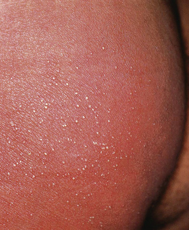
Fig. 7.9 Acute generalized exanthematous pustulosis. Courtesy, Yale Dermatology Residents’ Slide Collection. From Bolognia JL, Schaffer JV, Duncan KO, Ko CJ. Dermatology Essentials, 1e. Philadelphia: Saunders, 2014, with permission.
Related posts:


Stay updated, free articles. Join our Telegram channel

Full access? Get Clinical Tree


