Approximately 8% of males and 0.5% of females live with some form of Color Vision Deficiency (CVD). Medical professionals and surgical residents are not exempt from these statistics. While a mild red-green deficiency might only be a minor inconvenience in everyday life, in highly visual fields like plastic surgery and dermatology, the inability to distinguish subtle hues can compromise diagnostic accuracy and patient care.
This article explores the specific clinical challenges posed by CVD, differentiates between cosmetic tints and medically engineered optical lenses, and explains how color-correcting contact lenses serve as a vital occupational tool for affected practitioners.
The Impact of CVD on Surgical and Dermatological Diagnostics
In reconstructive surgery, visual color differentiation is a critical component of postoperative monitoring. Surgeons rely heavily on assessing skin color for early signs of flap failure. Identifying venous congestion (often presenting as a subtle bluish or purplish tint) or arterial insufficiency (presenting as pallor) requires acute color perception. For a surgeon with Protanomaly or Deuteranomaly, these early warning signs can blend into the surrounding tissue tones, delaying necessary intervention.
Dermatological assessments present similar challenges. Identifying erythema—the redness associated with infection, inflammation, or early-stage melanomas—is notoriously difficult for practitioners with red-green color overlap.
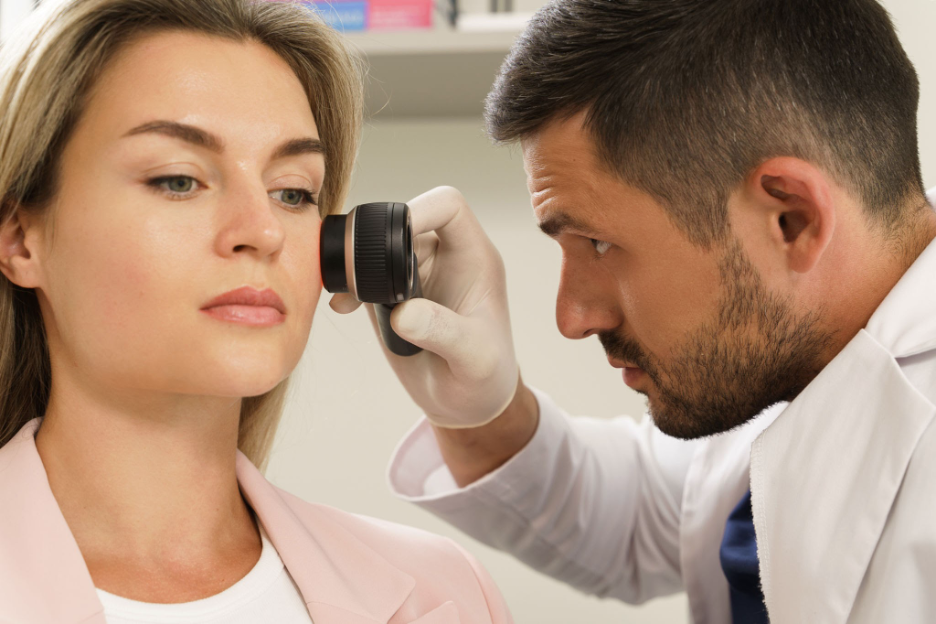
Assessing subtle skin erythema requires accurate color perception.. Source: Jun / Getty Images
When the visual contrast between healthy skin and inflamed tissue is muted, practitioners may be forced to rely more heavily on tactile feedback (like localized heat) or patient self-reporting, rather than immediate visual identification.
Mechanism of Action: Optical Filters vs. Cosmetic Tints
There is a common misconception in clinical settings that color-correcting contact lenses are akin to standard cosmetic colored contacts. Cosmetic lenses simply mask the natural iris with an opaque pigment to change eye color. Therapeutically engineered color-correcting lenses, however, function on an entirely different optical principle.
The biology of red-green CVD stems from a genetic variation that causes the chemical photopigments inside the red and green retinal cones to overlap in their light sensitivities. When a single wavelength of light triggers both cones simultaneously, the brain receives a confused signal, making it difficult to separate reds, greens, and browns.
Color-correcting contact lenses apply a precise, medically engineered magenta or red optical filter directly over the pupil. This filter acts as a targeted roadblock. It absorbs the specific wavelengths of light where the cone sensitivities overlap. By filtering out this “confusing” light, the lenses artificially force a sharper separation between the signals sent to the optic nerve. This drastically enhances contrast, allowing the clinician to perceive a wider, more distinct spectrum of color when examining tissue.
Why Contact Lenses Outperform Glasses in the O.R.
While colorblind glasses utilize similar wavelength-filtering technology, they are highly impractical in surgical or clinical settings. Glasses limit peripheral vision, can slide down the nose during long procedures, and often feature mirrored tints that are inappropriate for patient interactions.
More importantly, glasses interfere with clinical equipment. Because contact lenses sit directly on the cornea, they allow surgeons and dermatologists to seamlessly use surgical loupes, operating microscopes, dermatoscopes, and standard personal protective equipment (PPE) without any optical interference, reflection, or light leakage.
Screening and Self-Assessment for Medical Students and Residents
Despite the prevalence of CVD, there remains a stigma in medical education. Many medical students and residents hesitate to disclose color vision deficiencies due to fears of occupational limitation or being steered away from highly visual specialties.
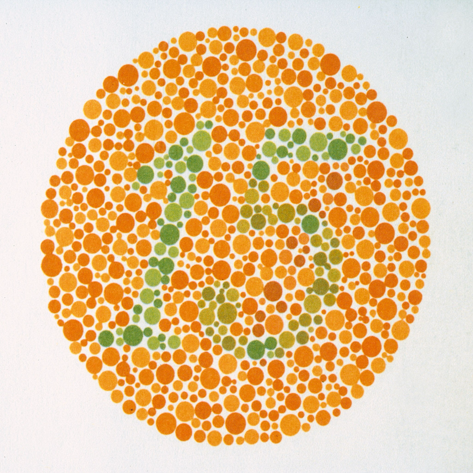
The Ishihara test remains a clinical standard for identifying red-green deficiencies.. Source: PHAS / Universal Images Group via Getty Images
Early and discreet self-assessment is critical. Before seeking a formal clinical diagnosis, students and practitioners can evaluate their baseline color perception using digital tools, such as the custom 24-plate interactive Ishihara vision test provided by optical platforms like Colorkinds. Identifying a deficiency early in one’s medical training allows the practitioner to proactively adopt optical aids before entering clinical rotations.
Conclusion
Color Vision Deficiency does not have to be a barrier to excellence in plastic surgery or dermatology. By understanding the underlying biology of retinal cone overlap and leveraging advanced optical filters, affected clinicians can restore diagnostic confidence. Incorporating color-correcting contact lenses into daily practice ensures that every practitioner, regardless of their baseline color perception, can deliver the highest standard of visual assessment and patient care.
Related posts:







Stay updated, free articles. Join our Telegram channel

Full access? Get Clinical Tree


