Fig. 9.1
Pneumoperitoneum
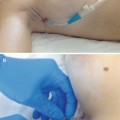
Fig. 9.2
Pneumoperitoneum done with a Veress needle. Three ports (one midline 10-mm port and two pararectal 5-mm ports) are inserted in the suprapubic region. The telescope is introduced into the 10-mm port
Fig. 9.3
Diagnostic laparoscopy and adhesiolysis
Fig. 9.4
The defect is made prominent by compression from outside
A 48-mm needle with double loop 1 (4 metric) 48 mm ½ circle heavy round bodied Ethilon (monofilament polyamide black) (Ethicon; Somerville, NJ, USA) is taken into the peritoneal cavity through a stab incision at the lower end of the diastasis. The needle is pulled inside by a needle holder and assisting grasper. Then the intracorporeal suturing is started (Fig. 9.5). When the suturing is completed, both edges have been approximated and a new linea alba constructed (Fig. 9.6, 9.7, and 9.8).
Fig. 9.5
Suturing of the edges of the defect is started with double loop 1 (4 metric) 48 mm ½ circle heavy round bodied Ethilon (monofilament polyamide black), creating a new linea alba
Fig. 9.6
Suturing in progress
Fig. 9.7
Suturing near completion
Fig. 9.8
Suturing completed
Then the 10-mm trocar is replaced by a 12-mm optical trocar, and the whole surgical team changes their gloves. An appropriate size of tissue-separating mesh is chosen. Prolene 2–0 sutures (Ethicon; Somerville, NJ, USA) are taken with long threads left on the upper, middle, and lower parts of the mesh in the midline, using all aseptic measures. Then the mesh is rolled like a cigarette, held with the needle holder, and taken into the abdominal cavity through the 12-mm optical trocar.
The mesh is unrolled and the pre-tied Prolene sutures are taken out transfascially with a suture passer in the midline; this technique reduces the chance of neural entrapment and postoperative pain. Then intracorporeal transfascial suture fixation of the mesh is performed with Prolene 2–0 (Figs. 9.9, 9.10, and 9.11
 Endoscopic Harvest of the Fascia Lata for Facial Reanimation
 Video-Assisted Thoracoscopic Sympathicotomies for the Treatment of Palmar and Axillary Hyperhidrosis
 Endoscope-Assisted Brow Lift
 Transaxillary Endoscopic Subfascial Breast Augmentation
 Endoscopic Flap Design and Harvesting
 Breast Reconstruction Using Laparoscopically Harvested Omental Flap


Related posts:
Endoscopic Harvest of the Fascia Lata for Facial Reanimation
Video-Assisted Thoracoscopic Sympathicotomies for the Treatment of Palmar and Axillary Hyperhidrosis
Endoscope-Assisted Brow Lift
Transaxillary Endoscopic Subfascial Breast Augmentation
Endoscopic Flap Design and Harvesting
Breast Reconstruction Using Laparoscopically Harvested Omental Flap
Stay updated, free articles. Join our Telegram channel
Full access? Get Clinical Tree
