Fig. 6.1
Dye injection into the subareolar region for sentinel lymph node biopsy
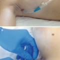
Next, the tumor is confirmed by means of 7.5-MHz ultrasonography, and a mixture of indigo carmine (12 mg) and xylocaine jelly 2 % (2–3 mL) (AstraZeneca; Tokyo, Japan) is injected to mark the resection line of the mammary gland at 12 point locations under ultrasound guidance, where the resection line is delineated further than 2.1 cm from the edge of the tumor. On the monitor, fluorescence images (Fig. 6.2) are obtained by using Photodynamic Eye (PDE) (Hamamatsu Photonics; Hamamatsu, SP, Japan), and subcutaneous lymphatic channels are usually detected over the skin within 1 or 2 min. When fluorescence images are poor, additional massage of the injection point is useful.
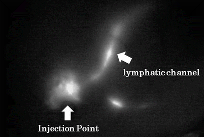
Fig. 6.2
Fluorescence images of the lymphatic channel on the monitor
The subcutaneous lymphatic channels are marked on the skin, based on the PDE image (Fig. 6.3).
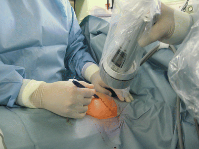
Fig. 6.3
Lymphatic channel marking on the skin based on Photodynamic Eye images
A 2-cm skin incision (Fig. 6.4) is made near the point of disappearance of the fluorescence under operating light conditions.
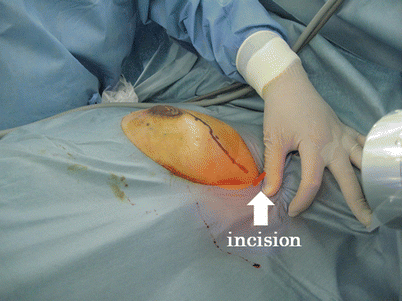
Fig. 6.4
Skin incision for sentinel lymph node biopsy
After skin incision, the subcutaneous fat tissues are dissected and the stained lymphatic channels are identified. Stained lymphatic channels are carefully dissected and traced until the first-drained lymph node is identified. The blue-stained SLNs are resected by direct visual inspection, together with the surrounding fatty tissue (Fig. 6.5). Usually, two or more lymph nodes are identified.
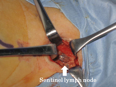
Fig. 6.5
Blue-stained sentinel lymph node
If blue-stained SLNs cannot be identified, the operating field is directly inspected using PDE, and the fluorescent lymph nodes are identified. Figure 6.6 illustrates a case in which the blue-stained lymph node could not be identified by direct visual inspection. Using the PDE, however, we were able to easily identify the SLN fluorescence on the monitor.
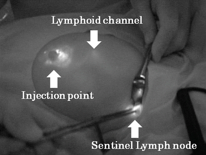
Fig. 6.6
Fluorescence image of operative field
Fluorescence imaging is also used for postoperative SLN confirmation, and EAPM is performed while the resected SLNs are pathologically examined by hematoxylin and eosin (HE) stain with frozen section.
The periareolar incision is placed at about 120°. From the incision, a Visiport™ Plus Optical Trocar (Covidien; Mansfield, MA, USA) with a rigid endoscope (Fig. 6.7) is inserted to create a 160°-wide subcutaneous tunnel. The light at the tip of the Visiport™ is used to confirm that the tip is inserted parallel to the skin and the breast tissue is being detached (Fig. 6.8).
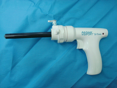
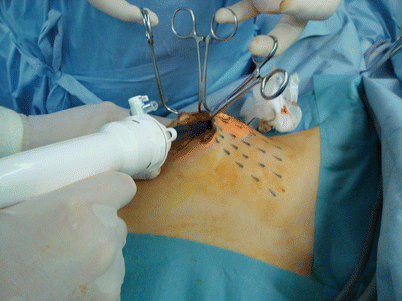
Fig. 6.7
Visiport™ optical trocar
Fig. 6.8
Detachment of subcutaneous tissue using Visiport™
Using PowerStar Bipolar Scissors (Ethicon; Somerville, NJ, USA) or Harmonic scalpel (Ethicon Endo-Surgery; Cincinnati, OH, USA), the tissue between the tunnels is cut (Fig. 6.9), and a skin flap with a slight amount of fat is created.
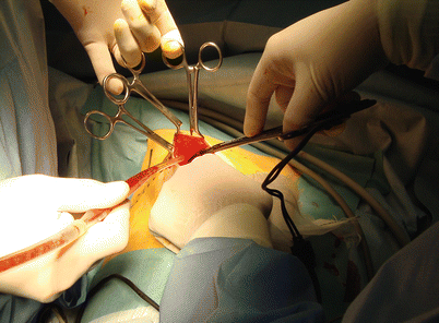
Fig. 6.9
Subcutaneous tunnel cutting using PowerStar Bipolar Scissors or Harmonic scalpel
A vertical incision is placed at the marking point closest to the nipple, using an electrocautery to expose and cut the pectoralis major fascia. Between the fascia and the muscle, a Round-Preperitoneal Distention Balloon (PDB) (Covidien; Mansfield, MA, USA) is inserted, and the mammary gland is separated from the pectoralis major muscle (Fig. 6.10). The balloon is inflated for 5 min (Fig. 6.11) and also serves for hemostasis.
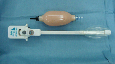
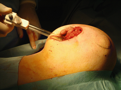
Fig. 6.10
Round-Preperitoneal Distention Balloon
Fig. 6.11
Inflation of the Preperitoneal Distention Balloon
At this stage, the direct-vision scope is inserted into the balloon. If the PDB is inserted properly, the muscle fibers of the pectoralis major muscle can be identified on the monitor.
The next step is excision of the tumor. The nipple is covered with gauzes to prevent it from being burnt, and the location of the tumor is reconfirmed by palpation. Using an electrocautery, the mammary gland is cut vertically while connecting the marking points, so that the excised tumor is a cylindrical mass (Fig. 6.12
 Endoscopic Harvest of the Fascia Lata for Facial Reanimation
 Video-Assisted Thoracoscopic Sympathicotomies for the Treatment of Palmar and Axillary Hyperhidrosis
 Endoscope-Assisted Brow Lift
 Transaxillary Endoscopic Subfascial Breast Augmentation
 Endoscopic Flap Design and Harvesting
 Breast Reconstruction Using Laparoscopically Harvested Omental Flap


Related posts:
Endoscopic Harvest of the Fascia Lata for Facial Reanimation
Video-Assisted Thoracoscopic Sympathicotomies for the Treatment of Palmar and Axillary Hyperhidrosis
Endoscope-Assisted Brow Lift
Transaxillary Endoscopic Subfascial Breast Augmentation
Endoscopic Flap Design and Harvesting
Breast Reconstruction Using Laparoscopically Harvested Omental Flap
Stay updated, free articles. Join our Telegram channel
Full access? Get Clinical Tree
