88
Enchondroma
Kevin D. Plancher and Michael Bothwell
History and Clinical Presentation
A 22-year-old woman with no prior hand injury presents after feeling a snap in her left ring finger while playing volleyball. The patient reports pain and swelling. She also reports pain with motion of the finger.
Physical Examination
The finger is tender to palpation. Range of motion is limited due to pain.
Diagnostic Studies
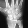
On radiographs a pathologic fracture in the hand or digits is frequently noted. Plain x-rays are diagnostic and reveal a centrally located lesion with marked bone expansion (Fig. 88–1). Cortical thinning and calcifications may also be present. Computed tomography (CT) has proven to be a useful tool in diagnosing enchondromas because it may detect endosteal scalloping and reveal the amount of bone destruction.
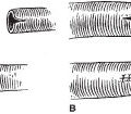
Histologically, this cartilage lesion is lobular and may penetrate the surrounding marrow spaces. The cells are often surrounded by a well-defined area of a proteoglycan matrix and a narrow rim of bone (Fig. 88–2).
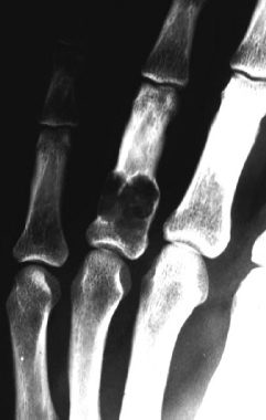
Figure 88–1. Radiographic finding of enchondroma of the proximal phalanx of the ring finger.
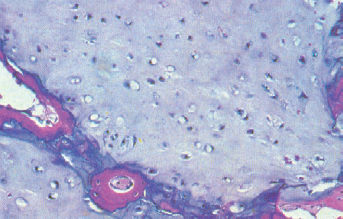
Figure 88–2. Histology of enchondroma showing cartilage lobular surrounded by narrow rim of bone.
Differential Diagnosis
Inclusion cyst
Fibrocystic defect of bone
Giant cell tumors
Related posts:
Stay updated, free articles. Join our Telegram channel

Full access? Get Clinical Tree


