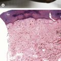
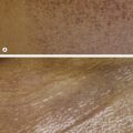
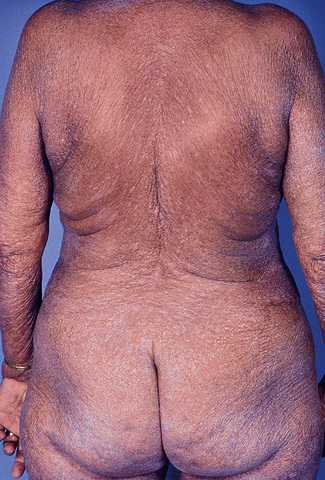
Fig. 3.4Sézary syndrome.Courtesy, Rein Willemze, MD. From Bolognia JL, Jorizzo JL, Schaffer JV. Dermatology, 3e. London: Saunders, 2012, with permission.
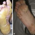
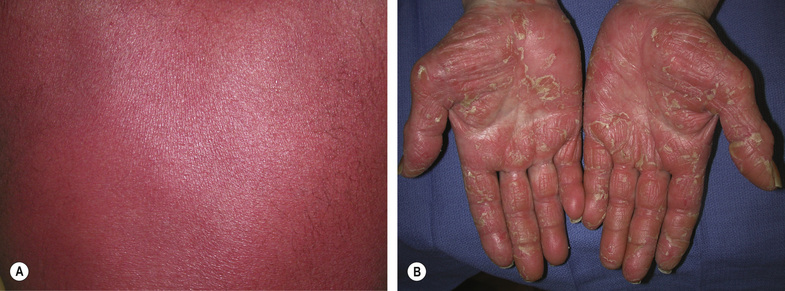
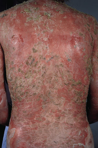
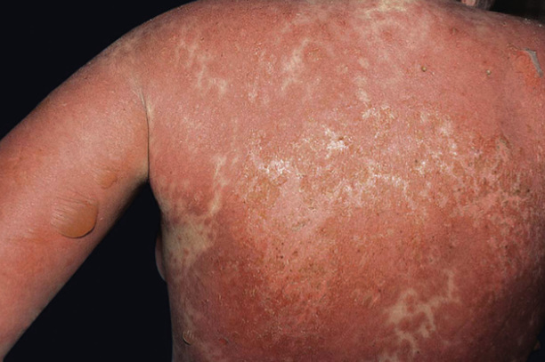
Atopic dermatitis and other eczematous processes as well as other diseases can also present with erythroderma (Figs 3.5–3.6).
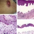
Fig. 3.5Pemphigus foliaceus.Courtesy, NYU Slide Collection. From Bolognia JL, Jorizzo JL, Schaffer JV. Dermatology, 3e. London: Saunders, 2012, with permission.
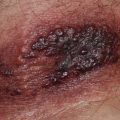
Fig. 3.6Toxic epidermal necrolysis-like presentation of acute lupus erythematosus (acute syndrome of apoptotic pan-epidermolysis [ASAP]).Courtesy, Yale Dermatology Residents’ Slide Collection. From Bolognia JL, Jorizzo JL, Schaffer JV. Dermatology, 3e. London: Saunders, 2012, with permission.
Photodistribution
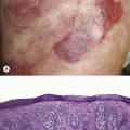
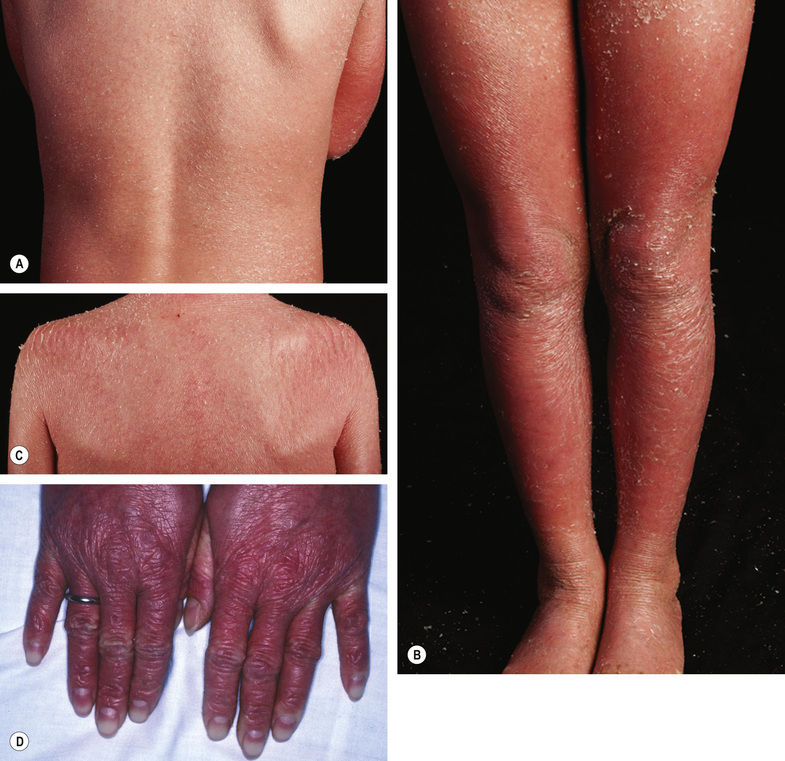
Once a photodistribution is determined (seeFig. 1.16A,Fig. 3.7), the primary involvement of the epidermis versus dermis and the morphology of primary lesions aid in narrowing the differential; for example, epidermal reactions including acute to chronic eczematous/spongiotic changes (Fig. 3.8), epidermal vesiculation (Fig. 3.9), erythematous papules and plaques (Fig. 3.10), and pigmented patches (Fig. 3.11).
Table 3.2
Photoreactions
Entity
Morphology
Histopathology
Epidermal photoreactions
Photoallergic reaction
• Acute spongiotic/eczematous process
• Vesicles
• Eosinophils
Phototoxic reaction (e.g. sunburn)
• Depending on severity, erythema to blistering
• Necrotic keratinocytes
Pellagra
• Flaky paint scale and erythema
• Parakeratosis and/or necrosis of the upper epidermis
Chronic actinic dermatitis
• Lichenification and erythema
• Mild spongiosis, acanthosis, hyperkeratosis
Hydroa vacciniforme
• Vesicles and erythema
Only gold members can continue reading. Log In or Register to continue


 Get Clinical Tree app for offline access