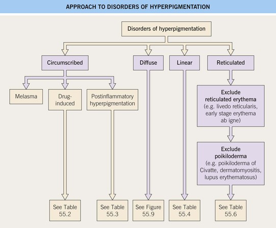
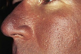
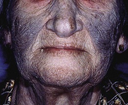
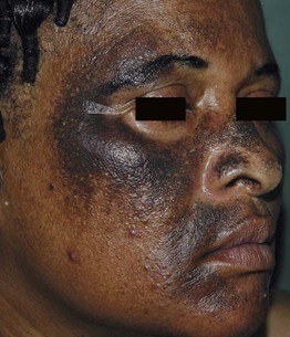
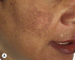
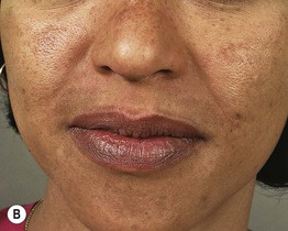
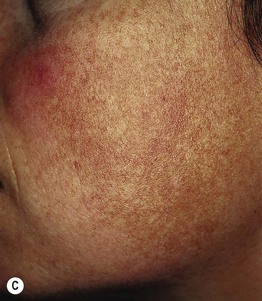
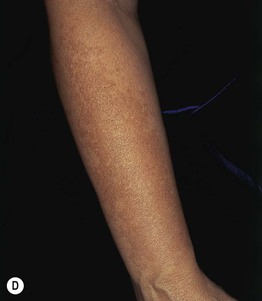
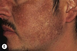
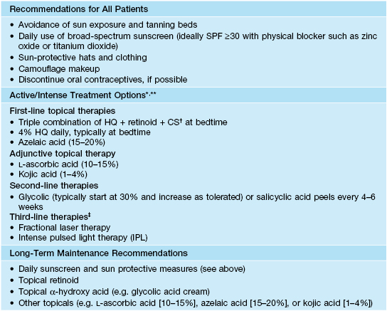
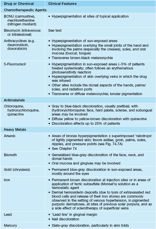
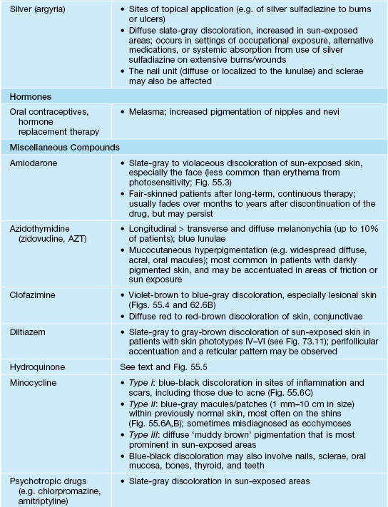
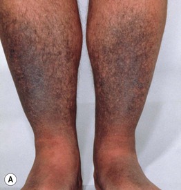
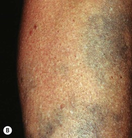
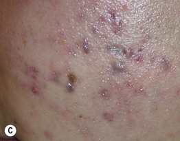
55 • Seen primarily in women; increased prevalence in individuals with skin phototypes III–IV. • Three classic clinical patterns based on distribution: centrofacial (most common), malar, and mandibular (Fig. 55.2). Fig. 55.2 Various forms of melasma and melasma-like hyperpigmentation. A Malar variant. B Mild centrofacial type with sparing of the philtrum. C Extension of the hyperpigmentation onto the mandible. D Involvement of the extensor forearm; note the same irregular outline as seen on the face. E Melasma-like appearance in a patient with previous acute cutaneous lupus erythematosus. C–E, Courtesy, Jean L. Bolognia, MD. • DDx: postinflammatory hyperpigmentation, drug-induced (e.g. minocycline, amiodarone), acquired bilateral nevus of Ota-like macules (especially in Asian women), actinic lichen planus, pigmented contact dermatitis, exogenous ochronosis due to the application of hydroquinone-containing bleaching agents, erythromelanosis faciei. • Treatment options are outlined in Table 55.1; typically epidermal hyperpigmentation responds best to treatment. Table 55.1 Suggested treatment options for melasma. * Results from topical treatments may take up to 6 months to appreciate; depending on the patient, HQ and combination HQ + retinoid + CS are typically used daily for 2–4 months and then decreased in frequency to 1–2 times per week; prolonged daily use can result in side effects such as perioral dermatitis, telangiectasias, and atrophy (CS) and ochronosis (HQ). ** While topical HQ can cause an allergic contact dermatitis, all topical agents may cause an irritant contact dermatitis, which can result in worsening of the dyspigmentation; if this is a concern, a small, nonfacial site test can be performed prior to widespread facial application. † Typically a Class 5–7 topical CS is used (see Appendix). ‡ Potential risk of post-procedural dyspigmentation; a site test should be performed prior to widespread facial laser or light therapy. HQ, hydroquinone. • The most common culprits of drug-induced circumscribed hyperpigmentation and discoloration are minocycline and the antimalarials (Table 55.2; Figs. 55.3–55.6). Table 55.2 Drugs and chemicals associated with circumscribed hyperpigmentation or discoloration. Diffuse hyperpigmentation and discoloration is discussed in Fig. 55.9. BCNU, 1,3-bis (2-chloroethyl)-1-nitrosourea. Fig. 55.6 Minocycline-induced discoloration. A The distribution on the shins and the gray-blue color can be similar to that seen with antimalarials. B Sometimes the discoloration is misdiagnosed as ecchymoses, but the subsequent color changes of green and yellow do not occur. C Blue-black pigmentation within acne scars and inflammatory papules. A, Courtesy, Mary Wu Chang, MD; C, Courtesy, Richard Antaya, MD. •
Disorders of Hyperpigmentation
Definitions
Approach to Disorders of Hyperpigmentation
Circumscribed Hyperpigmentation
Melasma
Drug-Induced Circumscribed Hyperpigmentation and Discoloration
![]()
Stay updated, free articles. Join our Telegram channel

Full access? Get Clinical Tree


