Dermatology for Plastic Surgeons II–Cutaneous Malignancies


Dermatology for Plastic Surgeons II–Cutaneous Malignancies


Daniel J. Ceradini

Keith M. Blechman


INTRODUCTION

Skin cancer is the most common form of cancer diagnosed in the United States, exceeding the combined incidence of breast, prostate, lung, and colon cancer cases annually. It is estimated that one in five Americans will be diagnosed with skin cancer during their lifetime.1 The incidence of cutaneous malignancies has increased dramatically over the past several decades. The most recent analysis of the incidence of non-melanoma skin cancer in the US population suggests that nearly 3.5 million new cases are diagnosed annually in 2 million patients.2 This figure has tripled from the estimated annual incidence of cutaneous malignancy from 1994 and signifies a major public health problem that accounts for over 2 billion dollars in healthcare spending annually. The treatment of skin cancer comprises over 75% of all procedures performed by plastic surgeons in the United States.3


BASAL CELL CARCINOMA


Epidemiology and Risk Factors

The vast majority of skin cancer cases diagnosed in the United States are either basal cell carcinoma (BCC) or squamous cell carcinoma. BCC is by far the most common cutaneous malignancy worldwide and accounts for nearly 80% of all skin cancers. There is marked worldwide geographic variability, most often affecting light-skinned populations in locations with the highest UV exposure. Although mortality is rare, locally aggressive BCC can result in significant patient morbidity.

The predominant risk factor for BCC is intense sunlight and UV exposure. Other risk factors include Fitzpatrick skin types I-II, a family history of skin cancer, male sex, smoking, human papilloma virus (HPV), exposure to arsenic or hydrocarbons, previous radiation, and immunodeficiency resulting from either acquired immunodeficiency syndrome or systemic drugs required for transplant recipients. While most arise sporadically, BCC is also associated with several clinical syndromes, including Bazex syndrome, Gorlin syndrome (basal cell nevus syndrome), and xeroderma pigmentosum. Constitutive activation of the sonic hedgehog signaling pathway is thought to play a significant role in BCC pathogenesis.


Diagnosis and Staging

Most BCCs arise in the head and neck and sun-exposed extremities and can be categorized into one of several different tumor types that exhibit distinct growth patterns. Tumor type is a key prognostic factor and guides selection of treatment options. Nodular BCC is the most common tumor type that classically presents as a dome-shaped nodular papule with a pearly surface, scattered telangiectasias, and rolled borders. As it enlarges, it typically ulcerates centrally, giving it the classic rodent ulcer appearance (Figure 14.1). Cystic BCC is a variation of the nodular type characterized by a cystic mucin-filled central core that retains the clinical appearance of the nodular tumors. Pigmented BCC is another variation of a nodular tumor that has brown or black macules, which can be easily confused with seborrheic keratosis or nodular malignant melanoma. Superficial BCC is the second most common tumor type. It appears as a demarcated multi-centric erythematous patch, frequently occurring on the trunk and extremities. The surface of this lesion is often scaly and ulcerated and can be misdiagnosed as a cutaneous fungal infection, discoid eczema, actinic keratosis, or psoriasis. Although there is frequently “normal” appearing skin between the tumor foci suggesting that each arises separately, this subtype exhibits a significant radial growth pattern and each focus is actually connected, likely arising from a single primary focus. Morpheaform or sclerosing BCC is the most aggressive tumor type, usually found in the head and neck. It is the most difficult type to diagnose and manage due to its insidious onset and infiltrative growth characteristics. It frequently appears as a poorly defined flat indurated plaque that resembles a scar without a history of trauma. Histologically, this tumor exhibits numerous thin linear extensions that can reach into the deep dermis, making surgical resection difficult and recurrence frequent.

Tissue biopsy definitively establishes the diagnosis and characterizes the histologic subtype. Additional workup is generally not required due to the very low rate of metastasis (<0.05%) and should be reserved for patients where a genetic disorder or clinical syndrome is suspected. The overall cure rate for BCC can exceed 90%; however, recurrence is frequent and more difficult to manage. The likelihood of curative treatment is determined by several prognostic factors, which allows clinicians to identify high-risk lesions that have an increased likelihood for recurrence of a more aggressive course (Table 14.1). Risk stratification guides selection of an appropriate treatment modality for tumor eradication. Staging of BCC is rarely performed due to the very low frequency of spread to lymph nodes or distant sites.


FIGURE 14.1. Nodular basal cell carcinoma. Central ulceration gives rise to the classic rodent ulcer appearance. (Right photo courtesy of Christine Liang, MD.)


TABLE 14.1 FEATURES OF HIGH-RISK BASAL CELL CARCINOMA AND CUTANEOUS SQUAMOUS CELL CARCINOMA


Anatomic site


Central face, periorbital region, ears/postauricular region, hands, feet


Tumor size


>5 mm on high-risk sites of central face, ears, hands, feet


>10 mm on scalp, forehead, cheek, neck


>20 mm on trunk, extremities


Histologic characteristics


Morpheaform/sclerosing, infiltrative, or micronodular subtypes (BCC)


Basosquamous differentiation


Perineural invasion


Clinical features


Recurrent tumors


Previously irradiated site


Poorly defined clinical borders


Immunosuppression


Site of chronic inflammation


BCC, basal cell carcinoma.


Surgical Management

The options for primary tumor eradication of BCC can be divided into destructive or surgical/excisional modalities. Destructive treatment options are generally reserved for lowrisk basal cell tumors and use a variety of methods to destroy neoplastic tissue including electrosurgery, cryosurgery, topical 5-fluorouracil, topical imiquimod, intralesional interferon, radiation, and photodynamic therapy. Notably, these modalities do not definitively ensure a margin clear of neoplasm. However, in selected low-risk cases, the overall success rate can be excellent.

Surgical or excisional treatment of BCC can be used in low-risk and high-risk cases. Simple excisional biopsy of low-risk lesions in anatomically simple areas (trunk and extremities) can result in success rates over 95%.4 In order to achieve histologically negative margins, guidelines exist to assist the surgeon: for tumors <1 cm a clinical margin of 4 to 5 mm, and for tumors >1 cm a clinical margin of 5 to 10 mm is recommended.5 In high-risk cases, especially on the face where obtaining adequate margins may result in significant deformity, direct excisional biopsy allows for histologic evaluation of the surgical margins to ensure that they are free of tumor to maximize the aesthetics of reconstruction. Mohs’ micrographic surgery is the most definitive modality and treatment of choice in high-risk BCC of anatomically complex areas on the face. High-risk tumors are serially excised and the entire margin of resection is examined histologically by the Mohs’ surgeon. Presence of tumor at the margins is mapped, and further excision of affected areas is serially performed and examined until the margins are clear. Mohs’ surgery achieves over 98% cure rate in primary tumors and over 95% in recurrent cases, although it is more expensive and time consuming to perform. The primary advantage of Mohs’ surgery is that it spares normal tissue and anatomic structures in complex areas while ensuring negative margins and excellent cure rates. Based on a recent randomized clinical trial comparing Mohs’ surgery to direct excision of BCC, Mohs’ surgery resulted in better outcomes for treatment of recurrent basal cell tumors, while there were no significant statistical differences in the treatment of primary BCC.6

BCC generally carries a good prognosis as the tumors tend to grow slowly and metastasize very rarely, but can result in significant morbidity due to local invasion. One-third of recurrences occur in the first year following treatment, half in the second year, and two-thirds in the third year regardless of the treatment modality.7 Patients should be monitored every 6 months for the first year following treatment and annually thereafter.


SQUAMOUS CELL CARCINOMA


Epidemiology and Risk Factors

Cutaneous squamous cell carcinoma (cSCC) is the second most common skin cancer, accounting for 15% to 20% of all cases of skin cancer in the United States. Similar to BCC, there is marked geographic variability in the incidence of cSCC, with more patients affected in areas with increased sun exposure. Although primary tumors can be locally invasive, it is frequently diagnosed in the early stages when it is a highly curable disease. Approximately 3,000 patients die from cSCC annually in the United States,8 and the incidence of more aggressive or advanced tumors is increasing.

Chronic cumulative sun exposure is the prevalent risk factor for cSCC, and both UVA and UVB are implicated in tumor pathogenesis. This is significant because the sun protection factor in sunscreens only measures protection against UVB. The incidence of cSCC increases significantly with age, likely reflecting an increased cumulative exposure to sunlight. Other environmental risk factors for cSCC include a history of radiation, chronic inflammation (as in Marjolin’s ulcer), and exposure to arsenic and hydrocarbons. Chronic immunosuppression secondary to organ transplantation markedly increases the risk of cSCC up to 250 times the general population and is closely correlated to the type of transplant, immunosuppressive drug burden, and time since transplantation.9,10 Host risk factors for cSCC include Fitzpatrick I-II skin types, fair hair, previous history of non-melanoma skin cancer, and infection with HPV. Additionally, certain inherited disorders such as xeroderma pigmentosum, epidermolysis bullosa, and albinism confer a genetic susceptibility to developing cSCC. UV-induced mutations in the p53 tumor suppressor gene are thought to be the molecular mechanism of malignant transformation of keratinocytes.


Diagnosis and Staging

The majority of cSCC is diagnosed on sun-exposed skin of the head and neck, dorsum of hands, lower arms, and legs. Unlike BCC, however, cSCC can arise from a premalignant actinic keratosis, identified as an area of erythematous, rough, scaly plaque that exhibits dysplastic growth and malignant potential. Up to 80% of cSCC tumors arise in association with a preexisting actinic keratosis, although overall <1% of all actinic keratoses undergo malignant transformation annually.11,12,13 Features of actinic keratosis that are associated with malignant transformation include inflammation, diameter >1 cm, rapid growth, ulceration, bleeding, and erythema.14 A cutaneous horn is a clinical variant of actinic keratosis that presents as a hyperkeratotic protuberance shaped like a cone extending above the plane of the skin. Approximately 15% of cutaneous horns actually contain cSCC,15 and excision is indicated.

cSCC in situ, also referred to as Bowen’s disease, frequently presents as a slowly growing, erythematous, scaly
patch. It is most frequently diagnosed in older patients (>60 years) and can occur anywhere on the body including the mucosal surfaces. When cSCC in situ occurs on the mucocutaneous epithelium of the glans of the penis or labia majora, it is referred to as erythroplasia of Queyrat. It occurs most often in uncircumcised men and is thought to be associated with chronic irritation, infection with HPV, and immunosuppression. It classically appears as a velvety red plaque on the glans of the penis. Progression to invasive cSCC occurs in up to 33% of cases over variable periods of time.16 When cSCC in situ occurs in the oral or genital mucosa, it presents as adherent white patches clinically referred to as leukoplakia. Notably, this must be differentiated from other causes of leukoplakia such as chronic irritation (usually from smoking), candidal infection, and HPV infection. This often requires a biopsy of suspicious lesions. Squamous cell carcinoma develops in 10% to 20% of all patients with leukoplakia.

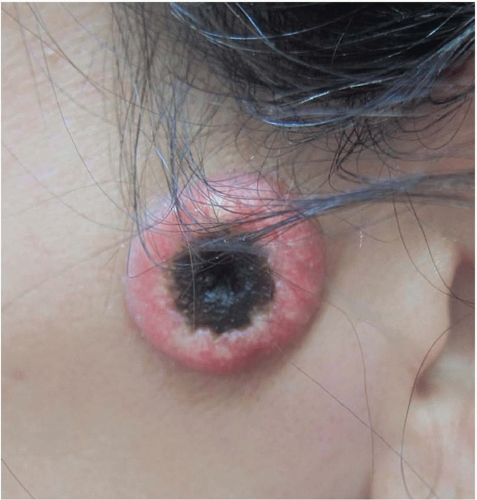
Keratoacanthoma is a rapidly growing nodule (over weeks to months) with a central ulceration or keratin plug that is found mainly in sun-exposed skin (Figure 14.2). Left untreated, it may spontaneously involute. Keratoacanthoma is felt to be a low-grade variant of cSCC, but is clinically difficult to distinguish from high-grade invasive cSCC. Shave biopsies are not helpful in making this distinction; therefore, surgical excision is recommended.

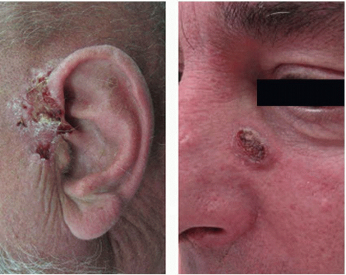
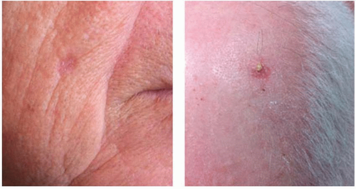
Invasive cSCC penetrates the basement membrane to reach the dermis and either arises de novo or is associated with actinic keratosis (Figure 14.3). Characteristic lesions are firm, raised, pink- or flesh-colored papules with frequent keratinization, scaling, ulceration, or crusting on the surface. These most often represent well-differentiated tumor types. Poorly differentiated lesions are typically soft, granulomatous nodules with areas of hemorrhage, necrosis, and ulceration and lacking in keratinization. Invasive cSCC associated with actinic keratosis in sun-exposed areas has a low metastatic risk and a favorable prognosis. De novo invasive cSCC, however, is a high-risk variant typically occurring in immunocompromised hosts or in areas of chronic irritation (such as burns) and has a metastatic rate as high as 14%.17


FIGURE 14.2. Keratoacanthoma with characteristic umbilicated center and keratin plug. If left untreated, it typically undergoes a period of rapid growth followed by spontaneous regression over several months; however, it can progress to squamous cell carcinoma with metastases.


FIGURE 14.3. Invasive cutaneous squamous cell carcinoma.

Diagnosis of cSCC is made by tissue biopsy to distinguish it from other neoplasms or cutaneous inflammatory conditions. In addition to definitive tumor diagnosis, patients with cSCC should undergo clinical examination of the appropriate draining lymph node basins. Palpable nodes should be biopsied by fine needle aspiration. Routine imaging studies for cSCC is not indicated, but should be obtained in patients that exhibit specific neurological symptoms or regional lymphadenopathy.

For the first time, the American Joint Committee on Cancer (AJCC) has introduced a completely separate staging system for cSCC, which was formerly incorporated into the “Carcinoma of the Skin” comprised of 80 different nonmelanoma skin cancers. This new TNM staging system utilizes a multidisciplinary evidence-based experience to more accurately describe the history and prognostic outcomes of cSCC (Tables 14.2 and 14.3).18 Due to the fact that most cSCC occurs in the head and neck, this system is meant to be consistent with the AJCC Head and Neck Staging system. The new cSCC staging system has several notable changes. The T staging (Tumor Characteristics) of the TNM system has been modified to eliminate the 5 cm size criteria and invasion of extradermal structures criteria to define a T4 lesion. Instead, a new list of “high-risk” features has been added, which impacts the overall T staging. Of these features, tumor grade now also contributes to the overall stage groups. The N component (Regional Lymph Nodes) of the TNM system has been totally revised to incorporate data indicating that overall survival decreases with increased node size and number involved.

While the majority of cSCC is diagnosed and cured in the early stages, the reported rate of regional metastasis ranges from 0.5% to 10%. While there is no consistent definition or stratification of what features of a primary tumor are considered “high risk” for regional spread, there are a number of tumor- and patient-specific characteristics that can be used to guide management. Tumor-specific features that are considered “high risk” include tumors located on the ears, lips, or within chronic wounds or scars, horizontal size >2 cm, thickness of 2 to 6 mm (low risk) or >6 mm (high risk), poorly differentiated cell types, perineural invasion, and rapidly growing or recurrent lesions.19 Patients who are organ transplant recipients or who are diagnosed with chronic lymphocytic leukemia, small lymphocytic lymphoma, epidermolysis bullosa, or HIV/AIDS are more likely to exhibit more aggressive tumor types and disease progression (Table 14.1).


Surgical Treatment

The surgical treatment options for cSCC are similar to those of BCC and are based on assessing the risk of local regional recurrence or distant metastasis. In selected low-risk cases, destructive treatment modalities can be used with excellent results. Direct surgical excision can be used for both lowrisk and high-risk lesions. In order to increase the chance of
achieving histologically negative margins, the recommended surgical margin for low-risk lesions is 4 mm, and for high-risk lesions it is 6 to 10 mm. An increasing number of high-risk features of the primary tumor may require a larger margin of resection. In anatomically complex areas of the face or in particularly high-risk cSCC tumors, Mohs’ micrographic surgery is the treatment of choice. Since cSCC tends to metastasize to the lymph nodes preferentially, there is some interest and initial success in using sentinel lymph node biopsy to diagnose subclinical lymph node metastasis and stage high-risk tumors. However, more controlled prospective randomized trials are required to determine whether detection of subclinical nodal metastasis will result in better clinical outcomes.20 There is currently no role for adjuvant therapy in patients who are at risk for recurrence. Patients with distant metastasis or advanced local disease not amenable to surgery or other treatment modalities require systemic chemotherapy.


TABLE 14.2 TNM STAGING CATEGORIES FOR CUTANEOUS SQUAMOUS CELL CARCINOMA


T CLASSIFICATION


TUMOR SIZE


Tis


Carcinoma in situ


T1


Tumor ≤2 cm in greatest dimension with <2 “high-risk” featuresa


T2


Tumor >2 cm in greatest dimension with or without one additional “high-risk” featurea or any size with ≥2 “high-risk” featuresa


T3


Tumor with invasion of maxilla, mandible, orbit, or temporal bone


T4


Tumor with invasion of skeleton (axial or appendicular) or perineural invasion of skull base


N CLASSIFICATION


NODAL METASTATIC BURDEN


N0


No regional lymph node metastasis


N1


Metastasis in single ipsilateral lymph node, ≤3 cm in greatest dimension


N2a


Metastasis in single ipsilateral lymph node, >3 but <=6 cm in greatest dimension


N2b


Metastasis in multiple ipsilateral lymph nodes, <=6 cm in greatest dimension


N2c


Metastasis in bilateral or contralateral lymph nodes, <=6 cm in greatest dimension


N3


Metastasis in lymph node, >6 cm in greatest dimension


M CLASSIFICATION


SITE


M0


No distant metastasis


M1


Distant metastasis present


a “High-risk” features include horizontal size >2 cm, depth ≥2 mm, perineural invasion, location on the ear, lip, or within a chronic wound or scar, poorly differentiated cell types, rapid growth, or recurrence.


Edge SB, Byrd DR, Compton CC, et al., eds. AJCC Cancer Staging Manual. 7th edn. New York, NY: Springer; 2010.


TABLE 14.3 ANATOMIC STAGE/PROGNOSTIC GROUPS FOR CUTANEOUS SQUAMOUS CELL CARCINOMA


STAGE


T


N


M


0


Tis


N0


M0


I


T1


N0


M0


II


T2


N0


M0


III


T3


N0 or N1


M0


T1 or T2


N1


M0


IV


T1, T2, or T3


N2


M0


Any T


N3


M0


T4


Any N


M0


Any T


Any N


M1


Reproduced from Farasat S, Yu SS, Neel VA, et al. A new American Joint Committee on Cancer Staging system for cutaneous squamous cell carcinoma: creation and rationale for inclusion of tumor (T) characteristics. J Am Acad Dermatol. January 2011;64:1051-1059.


MALIGNANT MELANOMA


Epidemiology and Risk Factors

Malignant melanoma is the most deadly form of skin cancer, diagnosed in 114,900 patients (46,770 noninvasive and 68,130 invasive) and resulting in 8,700 deaths in the United States in 2010.1 The incidence has been steadily increasing
over the past 50 years (currently 20 in 100,000 people), with notable growth in young white women 15 to 39 years (3% annual increase over the past 15 years) and white men over 65 years (5.1% annual increase since 1975). Overall, it is the fifth most common malignancy in men and the seventh in women, and 1 in 52 people will be diagnosed with melanoma in their lifetime. Although melanoma accounts for less than 5% of all skin cancer cases, it results in greater than 75% of skin cancer deaths–approximately one person dies of melanoma every hour. Indeed, melanoma is a significant public healthcare problem–the incidence is increasing, there is a predominant modifiable risk factor (sun exposure), and it is curable in the very early stages but not in advanced disease.

Assessment of risk factors for melanoma can be divided into host factors and environmental factors. Host factors that confer a higher risk of developing melanoma include physical attributes such as fair features, Fitzpatrick I-II skin types, and blue/green eyes. Additionally, numerous common congenital nevi, atypical nevi, and giant nevi are all associated with increased risk. A personal history of melanoma is thought to confer a 5% lifetime risk of developing a second melanoma. Finally, familial melanoma accounts for approximately 10% of all cases and is associated with mutations within the cyclin-dependent kinase inhibitor 2A locus (CDKN2A), cyclin-dependent kinase 4 (CDK4), and melanocortin 1 receptor (MC1R).

Perhaps the most significant and modifiable environmental risk factor for melanoma is sun exposure, particularly intermittent and intense exposure. UVA and UVB exposure are both strongly associated with melanoma. One or more blistering sunburns early in life or greater than five sunburns at any age increases the lifetime risk of developing melanoma twofold. Regular broad spectrum sunscreen use may reduce the risk of developing invasive melanoma.21 It is important to note that while intense sun damage is very strongly associated with melanoma, it is not necessarily required for malignant transformation, as a significant number of lesions arise in relatively sun-protected areas (soles of feet, anus, and vagina). This underscores the complex multifactorial role of host and environmental factors in melanoma pathogenesis.


Diagnosis and Staging

Since early detection of melanoma is critical to improve public healthcare outcomes, the simple yet effective ABCDE diagnostic tool was developed at the New York University Langone Medical Center to educate the public and general healthcare practitioners, simplifying the decision to biopsy a suspicious lesion (Figure 14.4).22 This tool utilizes five simple criteria for identifying pigmented lesions that are suspicious for melanoma: Asymmetry, Border Irregularity, Color Variegation, Diameter > 6 mm, and Evolution or change in the appearance of lesion over time. Using these criteria, the vast majority of melanomas are detected by clinical examination. It is important to note, however, that a minority of lesions are atypical and can be nonpigmented (5%), resemble other types of cutaneous malignancies (basal or squamous cell carcinoma), or be smaller than 6 mm in size.


FIGURE 14.4. Malignant melanoma. Lesions suspicious for melanoma demonstrate asymmetry, border irregularity, color variegation, diameter >6 mm, and evolution or change in the appearance. (Photos courtesy of Richard L. Shapiro, MD.)

Melanoma can be classified into five clinical and histologic growth patterns, each of which has unique clinical characteristics: superficial spreading, nodular, lentigo maligna, acral lentiginous, and desmoplastic melanoma. With the exception of nodular melanoma, the remaining subtypes originate from an in situ radial growth phase that does not have metastatic capability. The prognostic significance of growth pattern remains controversial, although there are some genetic factors identified within the subtypes that may have prognostic value. Superficial spreading melanoma presents as flat or slightly elevated lesion with variegate pigmentation, most commonly occurring on the trunk in men and the legs in women, in patients aged 30 to 50 years. As the name would imply, the growth pattern is typically superficial and radial with scattered atypical melanocytes in the epidermis. It is the most common subtype in the Caucasian population and likely contributes significantly to the increasing incidence of melanoma over the past 30 years. Nodular melanoma is the second most common growth pattern, which often lacks the classic features commonly identified by the ABCDE melanoma screening tool. These lesions commonly present as a smooth, single colored (black or brown) elevated nodule or an ulcerated mass on examination, frequently affecting the legs or trunk. They are typically thicker and more advanced at the time of diagnosis, largely due to a relatively short or lack of radial growth phase. Overall, nodular melanomas account for most thick melanomas; however, the survival rate and prognosis is similar to that of the other clinical types when thickness and ulceration are taken into account. Both nodular and superficial spreading melanomas are associated with increased sun exposure in fair-skinned individuals. Lentigo maligna melanoma (Figure 14.5) is a slow growing lesion with radial spreading that typically arises in longstanding pigmented lesion on chronically sun-damaged anatomic sites (head and arms) in older patients. Hypopigmented lesions are also possible within this subtype. It occurs most often in fair-skinned older individuals, with an average age of diagnosis at 65 years, and is associated with solar elastosis of the surrounding skin. Acral lentiginous melanoma affects only 2% to 8% of Caucasians, but accounts for up to 36% of melanoma diagnosed in African Americans, making it the most common subtype within this demographic.23 It commonly occurs on the palms of the hand, sole of the feet, or beneath the nail plate (subungual) and presents at a more advanced stage with an aggressive course compared with the other subtypes. Subungual variants commonly present as a longitudinal line of pigment extending the length of the nail plate, with the hallmark spread of the pigment to the proximal nail fold referred to as Hutchinson sign. Finally, desmoplastic melanoma is a relatively uncommon subtype that presents as an unremarkable plaque or nodule and can easily be misdiagnosed at an early stage. It affects older patients (although not as old as lentigo maligna melanoma) most commonly in the head and neck and occurs in men twice as often as in women. Desmoplastic melanoma is frequently associated with nerve invasion and spread along fascial planes and tends to be thicker at the time of diagnosis. They are locally aggressive with a higher rate of local recurrence, but exhibit a low incidence of lymph node involvement. These clinical features more closely resemble a soft tissue sarcoma, suggesting that the underlying biology of desmoplastic melanoma may be unique compared with the other subtypes.

Only gold members can continue reading. Log In or Register to continue

Stay updated, free articles. Join our Telegram channel

Jun 26, 2016 | Posted by in General Surgery | Comments Off on Dermatology for Plastic Surgeons II–Cutaneous Malignancies

Full access? Get Clinical Tree

Get Clinical Tree app for offline access