Fig. 21.1Common sites of cysts (A), and developmental anomalies of the face and neck (B).B, From Bolognia JL, Jorizzo JL, Schaffer JV. Dermatology, 3e. London: Saunders, 2012, with permission.
Fig. 21.2Epidermoid cyst. A central punctum (pore) is often evident. Wall resembles normal epidermis with central flaky keratin.
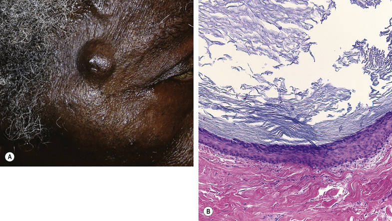
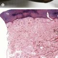
Fig. 21.3Pilar cyst. This cyst is most common on the scalp. Wall lacks a granular layer and surrounds dense keratin.A, Courtesy, Mary Stone, MD. A, From Bolognia JL, Schaffer JV, Duncan KO, Ko CJ. Dermatology Essentials, 1e. Philadelphia: Saunders, 2014, with permission.
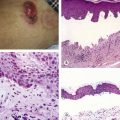
Fig. 21.4Hidrocystoma. Translucent to blue papule, often on the eyelid margin. Double-layered epithelium with “snouts” on the cell surface at lumen.
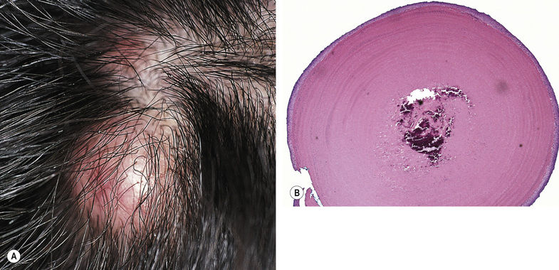
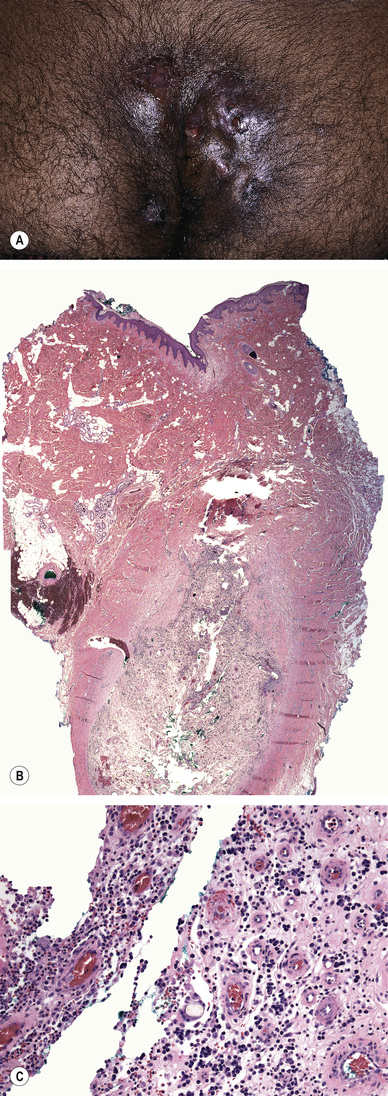
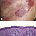
Fig. 21.5Pilonidal sinus tracts. Biopsy findings include epithelial lined tracts (not shown), acute and chronic inflammation, and free hair shafts. The most common location is the sacral area.Courtesy, Kalman Watsky, MD.
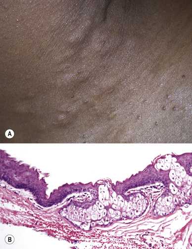
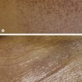
Fig. 21.6Steatocystoma multiplex. This cyst can develop on any site; when multiple, lesions are typically larger than vellus hair cysts and present on the trunk. Wall with sebaceous glands and an inner rim that is bright pink and undulating.A, Courtesy, Yale Dermatology Residents’ Slide Collection.
Only gold members can continue reading. Log In or Register to continue