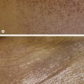
Histopathology:
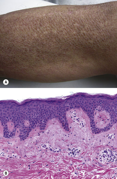
Light pink, smooth deposits in the papillary dermis, often with associated pigment incontinence; deposits are larger and associated with epidermal hyperplasia in lichen amyloidosis
Deposits stain with cytokeratins
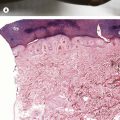
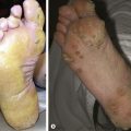
Pretibial Myxedema (Fig. 22.3)
Pink plaque(s) that are nodular in later stages, especially on the lower extremity
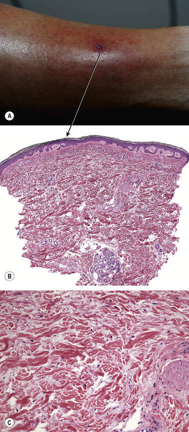
Histopathology:
Mucin throughout the reticular dermis
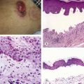
Adult Colloid Milium (Fig. 22.4)
Brown to yellow translucent papules, especially on chronically sun-damaged skin of the face and/or hands but other sites may be affected
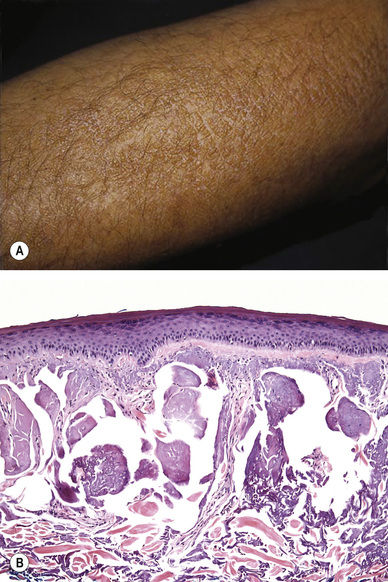
Histopathology:
Nodules of pink, fissured material
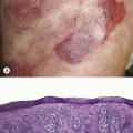
Lipoid Proteinosis (Fig. 22.5)
Genodermatosis with mutations in ECM1
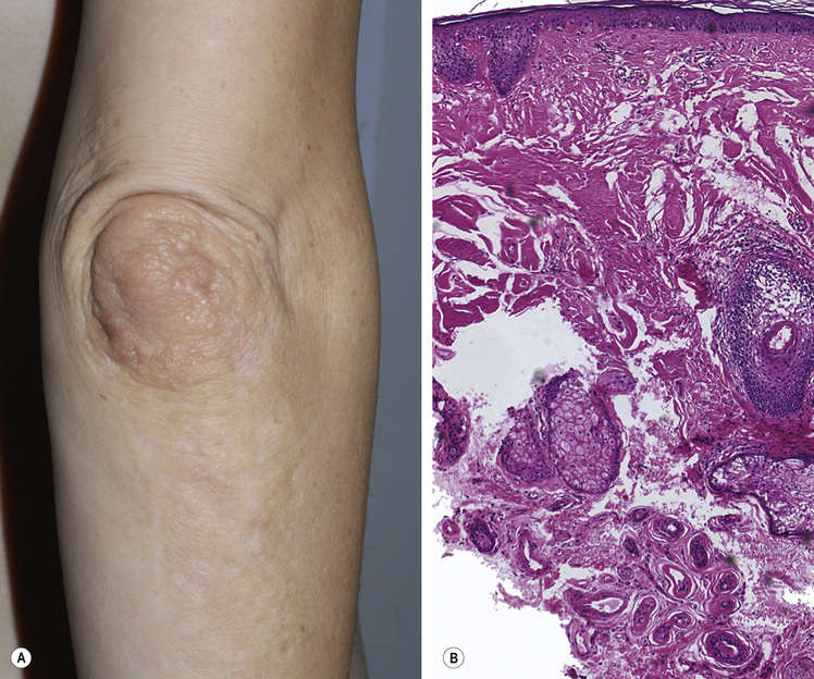
Waxy papules and plaques on extensor surfaces and face with scarring
Histopathology:
Pink dermal material, sometimes with a vertical orientation in the upper dermis; material may be accentuated around adnexal structures
Erythropoietic Protoporphyria (Fig. 22.6)
Genodermatosis, mutation in ferrochelatase
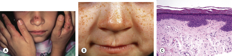
Sun-exposed skin is affected, especially the nose, cheeks, and dorsal hands
Erythema, erosions, waxy scarring
Histopathology:
Pink dermal material around vessels (early lesions) and/or filling the upper dermis (later lesions)
Related posts:

Stay updated, free articles. Join our Telegram channel

Full access? Get Clinical Tree


