Deltopectoral Skin Flap: Pharyngoesophageal Reconstruction
V. Y. BAKAMJIAN
Reconstruction of the pharynx and cervical esophagus was the initial purpose for which the deltopectoral flap was devised in 1962 to overcome the considerable difficulties and shortcomings of earlier methods, mainly the two-stage technique with a cervical flap (see Chapter 215) and other one-stage techniques with skin grafts (1, 2, 3, 4, 5, 6, 7).
OPERATIVE TECHNIQUE
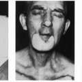
Radical neck dissection with laryngopharyngectomy that precedes reconstruction with a deltopectoral flap is best served by a transverse pair of parallel neck incisions, modified after MacFee. Contrary to commonly voiced opinions, these incisions amply provide needed exposure for accomplishing the ablative objective adequately (Fig. 216.1A) while also serving the interest of the reconstruction. The upper incision is placed at hyoid or just below hyoid level in a natural skin crease, and the lower is placed at clavicular level, in common with the upper margin of the deltopectoral flap (Fig. 216.1B).
The flap is introduced to the midline of the neck, passing beneath the bipedicle flap of cervical skin raised between the incisions of the neck dissection (Fig. 216.1C). With its raw side facing the prevertebral fascia, suturing begins between the tip of the flap and the excision line in the posterior wall of the oropharynx or epipharynx, as the case may be. The process continues to the sides bilaterally and reflects forward to meet at a point at the base of the tongue, a bit off-center and away from the side of the radical neck dissection. Starting from this point, the longitudinal seam that entubes the flap veers gently backward in its descent to meet the stump of the esophagus behind the transected trachea. Here the lips of the seam part to encircle the esophageal end opening, establishing an end-to-side anastomosis to the skin tube (Fig. 216.1D). Before this anastomosis is completed, however, a nasally introduced feeding tube is passed down the skin tube and into the esophagus and stomach, after which the seam is continued a short distance more to a fistulous exit over the medial head of the clavicle. The neck wounds are closed, and the donor wound on the chest and shoulder is covered with a skin graft (Fig. 216.1E).
Related posts:


Stay updated, free articles. Join our Telegram channel

Full access? Get Clinical Tree


