Decubitus ulcers


Decubitus ulcers


Joseph A. Witkowski, Lawrence Charles Parish, Caren Campbell and Jennifer L. Parish
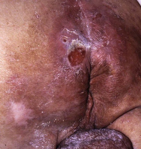

Evidence Levels:  A Double-blind study  B Clinical trial ≥ 20 subjects  C Clinical trial < 20 subjects  D Series ≥ 5 subjects  E Anecdotal case reports


image


The decubitus ulcer represents a defect in the skin that can extend through the subcutaneous tissue and muscle layer onto the underlying bone.


Management strategy


Prevention


A patient in an ordinary bed who is at risk of developing a decubitus ulcer, also referred to as pressure ulcer or bed sore, should be repositioned at frequent intervals; however, the correct timing for turning has never been established. The regularity is determined by the level of risk of developing an ulcer and the duration of blanchable erythema. Pillows and foam wedges are used to maintain position and to keep bony prominences apart. Completely immobile patients should have their heels raised from the bed by a pillow or boot and not be placed on their trochanters, unless a specialized bed is used. To avoid the latter, use a 30° position from the horizontal lying on the side. The head of the bed should not be raised more than 30° from the horizontal for an extended period. Where possible, use lifting devices or draw sheets to reposition or to transfer patients. Finally, when appropriate, the patient should be placed on a pressure-reducing device such as a foam, alternating air, gel, or water mattress.


A patient sitting in a wheelchair who is at risk for developing a decubitus ulcer should be repositioned frequently, perhaps every hour, and be taught to shift weight every 15 minutes. The chair should be adjusted appropriately for each patient. A pressure-reducing device made of foam, gel, air, or a combination of each is indicated. Unfortunately, even in paraplegics the exact time interval for repositioning has never been accurately determined.


Theoretically, decubitus ulcers should be preventable, but despite these measures, many simply cannot be prevented. If they are associated with immobility, sustained pressure, and the loss of pain sensibility, then these problems can and should be addressed. In practice, successful prevention is often foiled by our limited understanding of the pathogenesis, as well as by complicating comorbidities. There is also some evidence that many deep ulcers are initiated by multiple microthromboses of deep tissues. This indicates that dehydration, along with any factor that might increase blood coagulability, should be addressed.


Management


The management of skin lesions caused by pressure is based on four principles:


The patient should not lie on the ulcer. A patient who is at risk for developing additional ulcers and can assume a variety of positions without lying on the ulcer should be placed on a static support surface, i.e., air, foam, or water. If the patient cannot assume various positions without lying on the ulcer or bottoms out while on a static surface, or if the ulcer does not heal after 2 to 4 weeks of optimal care, place the patient on a dynamic support surface when possible, i.e., an alternating air overlay on the mattress, a low-air-loss bed, or an air-fluidized bed. If a patient has large deep ulcers (stage III or IV) on multiple sleep surfaces or has excess moisture on intact skin, use a low-air-loss bed or an air-fluidized bed. A patient with an ulcer on the sitting surface should not sit, if possible.


Removal of necrotic debris


Surgical debridement is indicated for infected ulcers with necrotic debris and eschars other than those on the heel; however, the extent of tissue needing to be removed is highly variable. An eschar on the heel should be excised only if it is fluctuant, draining, or surrounded by cellulitis, and if the patient is septic.


Major debridement is performed in the operating room, but serial sharp debridement can be performed at the bedside. The use of systemic antimicrobials should be considered to prevent bacteremia during significant debridement. A bone biopsy is recommended while debriding ulcers when bone is exposed and for non-healing deep ulcers (stage III or IV ulcers) after 2 to 4 weeks of optimal therapy.


Other ulcers can be debrided by the use of saline wet-to-dry gauze every 4 to 6 hours by the use of saline in a 35 mL syringe with an attached 19-gauge angiocatheter, or by whirlpool use. The use of enzymes should be reserved for ulcers that are not clinically infected. Autolytic debridement is indicated for non-infected ulcers that are not likely to become infected.


Debridement can also be indicated for staging of the ulcer. This assumes that staging is a requisite for treating the patient. Although debridement is a useful therapeutic tool, complete elimination of necrotic tissue is unnecessary, as is daily surgical debridement.


General measures


Treatment of the decubitus ulcer can be simplified and made more effective if the following recommendations are considered.


image Saline should be used to clean most pressure lesions; soap and disinfectants are too irritating for more than occasional use.


image When ulcers are not infected, synthetic dressings should be changed only if they become dislodged or wound fluid escapes from under the dressing.


image Periulcer skin must be kept dry not only to avoid maceration but also to permit the dressing to adhere to the skin.


image To obliterate dead space, fill deep ulcers loosely with a hydrocolloid, a hydrogel wound filler, or an alginate rope before applying a synthetic dressing. This same material should be placed under the edge of the ulcer when undermining is present. Bleeding after serial surgical debridement can often be controlled with an alginate dressing; the calcium alginate assists in the clotting pathway. Moistening with saline can loosen an alginate dressing that adheres to granulation tissue.


image A clean ulcer failing to show signs of healing, or an ulcer with persistent excessive exudate, should be treated with antibacterial agents, e.g., 1% silver sulfadiazine, cadexomer iodine, triple antibiotic, or retapamulin, for 2 weeks to reduce the bacterial burden. Increased bacterial burden may impede healing before clinical signs of infection become apparent. The odor of an infected ulcer can often be eliminated by applying metronidazole gel to the ulcer bed. Systemic antimicrobial agents are indicated for patients with bacteremia, sepsis, advancing cellulitis, or osteomyelitis.


image Although most synthetic dressings relieve pain, treatment for moderate to severe pain can include topical anesthetics, non-steroidal anti-inflammatory drugs (NSAIDs), opiates, antidepressants, and sedatives. Many patients with decubitus ulcers do not have pain.


Superficial and deep ulcers


Superficial and deep ulcers without necrotic debris are treated with saline wet-to-dry gauze or an adherent synthetic dressing. Deep ulcers should be loosely filled with a synthetic wound filler before applying a synthetic dressing. The deep ulcer with necrotic debris requires debridement and is then treated as a clean ulcer.


Enzymatic debridement or the use of an antimetabolite can help manage the eschar. Covering the lesion with an adhesive occlusive dressing for several days will often soften the eschar before excision is undertaken. Faster softening can be accomplished by scarifying the lesion, applying an enzyme to the surface, and covering with an impermeable plastic wrap. The firmly adherent dry eschar that is not attached to underlying bone can often be separated from the surrounding skin with 5% 5-fluorouracil cream. After scarification and application of zinc oxide paste to protect the surrounding skin, 5-fluorouracil is applied to the eschar, including its margin, and then covered with an impermeable plastic wrap. Application is repeated every 8 hours. When separation occurs, it can be excised.


Aug 7, 2016 | Posted by in Dermatology | Comments Off on Decubitus ulcers

Full access? Get Clinical Tree

Get Clinical Tree app for offline access