Craniofacial Clefts and Hypertelorbitism


Craniofacial Clefts and Hypertelorbitism


James P. Bradley

Henry K. Kawamoto


Congenital craniofacial clefts are malformations of the cranium and face with deficiencies or excesses of tissue along an anatomic line based on embryologic maldevelopment.1 They are among the most disfiguring of all facial anomalies. Craniofacial clefts exist in a multitude of patterns and varying degrees of severity.2 They are expressed either unilaterally or bilaterally. In addition, one cleft type may manifest on one side of the face, while a different cleft type is present on the other side.3


TESSIER CLASSIFICATION

The Tessier classification for rare craniofacial clefts is the most complete and has withstood the test of time.1 This classification links clinical observations with underlying skeletal deformities seen with preoperative 3D computed tomography (CT) scan imaging and confirmed during surgery. Newer neuroembryologic theories that allow for mapping of developmental zones of the face have confirmed the value of the Tessier classification to embryologists and geneticists, not just surgeons (Figure 26.1).4

In the Tessier classification, clefts are numbered from 0 to 14 (Figure 26.2). The eyelids and orbits are designated as the horizontal axis of this functional system dividing the face into upper and lower hemispheres. The orbit separates the facial clefts (0 through 7; going from medial to lateral) from the cranial clefts (8 through 14; going from lateral to medial). In many cases, the facial clefts extend into the cranium in predictable lines to form combination cranial and facial clefts, including 0 and 14, 1 and 13, 2 and 12, 3 and 11, 4 and 10, 5 and 9, and 6 and 8. The soft tissue and skeletal components are seldom affected to the same extent. The skeletal landmarks tend to be more constant and reliable than the soft tissue landmarks.


FIGURE 26.1. Embryological correlation to Tessier craniofacial clefts: Tessier-numbered craniofacial clefts are depicted as growth center junctions in this 45-day-old fetus.


DEFINING FEATURES OF CRANIOFACIAL CLEFTS


Number 0 Cleft

The number 0 cleft has been called median craniofacial dysraphia, centrofacial microsomia, frontonasal dysplasia, or median cleft face syndrome—but for accuracy it is the facial manifestation or lower half of “median craniofacial dysplasia.” Patients with this midline facial cleft may have a cranial extension or a number 14 cleft. The number 0 Tessier craniofacial clefts are unique in that there may be deficient, normal, or excess tissue. Tissue agenesis and holoprosencephaly (the hypoplasias) are one end of the spectrum, and frontonasal hyperplasia and excessive tissue (the hyperplasias) are the other end. Median anomalies with normal tissue volume occupy the middle portion of the spectrum (Table 26.1).5

Median craniofacial hypoplasia (deficiency of midline structures): A deficiency may manifest as hypoplasia or agenesis in which portions of midline facial structures are missing. This developmental arrest may range from the mildest form of hypoplasia of the nasomaxillary region and hypotelorism to a severe form of cyclopia, ethmocephaly, or cebocephaly. The subcategories in Table 26.1 demonstrate that the severity of the facial anomalies generally correlate with the severity of brain abnormality and mental retardation. Clinically, it may be important to distinguish among patients with poor brain differentiation (alobar holoprosencephaly) who may die in infancy from those with a better prognosis (lobar brain).


FIGURE 26.2. Tessier’s classification of craniofacial clefts: The left half (right side of the face) depicts the skeletal locations of numeric clefts and the right half (left side of the face) outlines the clinical locations of clefts based on soft tissue landmarks. Facial clefts, number 0 through number 7; cranial clefts, number 8 through number 14. Mandibular midline facial cleft number 30 is also seen.


TABLE 26.1 NUMBER 0 CLEFT: MEDIAN CRANIOFACIAL DYSPLASIA


I.


Median Craniofacial Hypoplasia (tissue deficiency or agenesis)


A.


Holoprosencephalic spectrum (Alobar Brain)


1.


Cyclopia: Single eye in a single orbit. Arhinia with proboscis often located above the single orbit and microcephaly.


2.


Ethmocephaly: Severe hypotelorism but separate orbits. Arhinia with proboscis located in between the orbits.


3.


Cebocephaly: Severe hypotelorism. Proboscis-like rudimentary nose.


4.


Agenesis of the primary palate and associated midline structures with severe hypotelorism.


B.


Median cerebrofacial hypoplasia (lobar brain): With midline facial hypoplasia and midline cerebral malformations. Unilateral or bilateral cleft lip and palate can be present.


C.


Median facial hypoplasia (lobar brain): Midline facial hypoplasia without gross cerebral involvement. Unilateral or bilateral cleft lip and palate can be present.


1.


Median facial hypoplasia full form


2.


Microform median facial hypoplasia


a. Binder syndrome


b. Central maxillary incisor anomaly


c. Absent upper lip frenulum


II.


Median Craniofacial Dysraphia (normal tissue volume but clefted)


A.


True Median Cleft:


It can be presented as isolated cleft of the upper lip “0 cleft” or with tissue deficiency or agenesis, “e.g., absent nasal septum.”


B.


Anterior Encephaloceles: An encephalocele is a cystic congenital malformation in which central nervous system structures herniated through.


III.


Median Craniofacial Hyperplasia (tissue excess or duplication)


This spectrum of anomalies include all forms of excess tissue starting from just thickened or duplicated nasal septum to the more severe forms of frontonasal dysplasia.


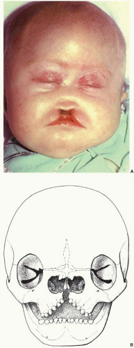
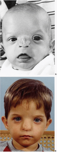
Soft tissue deficiencies with Tessier 0 clefts include the upper lip and nose. Agenesis or hypoplasia may result in a false median cleft lip and absence of philtral columns. When a wide central cleft exists, it typically extends the length of the upper lip and into the nasal floor (Figure 26.3A). With nasal anomalies the columella may be narrowed or totally absent. The nasal tip may be depressed from lack of septal support. The septum may often be vestigial with no caudal attachment to the palate. Dental abnormalities may include a single maxillary central incisor or even absent central maxillary incisors.

Skeletal deficiencies range from separation between the upper central canines to absence of the premaxilla and a cleft of the secondary palate (Figure 26.3B). Nasal deficiency may include partial or total absence of the septal cartilage and even nasal bones. The bone defect may extend cephalad into the area of the ethmoid sinuses and result in hypotelorism or cyclopia.


FIGURE 26.3. Number 0 cleft: Median craniofacial hypoplasia. A. Patient with midline facial hypoplasia. B. Illustration shows skeletal involvement with separation between the central incisors and widening of the nasal region and orbital hypertelorism.


Median craniofacial dysraphia (normal tissue volume but clefted): These Tessier 0 clefts have normal tissue volume but are abnormally split (true median cleft lip) or displaced (encephalocele).

Soft tissue involvement: When an isolated cleft of the upper lip is not associated with tissue deficiency (e.g., absent nasal septum) or tissue excess (e.g., duplicated septum), it is considered a “true” median cleft lip (Figure 26.4). With a true median cleft lip there is a split between the median globular processes; whereas, with a false median cleft lip an agenesis of the globular processes may occur.

Skeletal involvement: When the true median cleft passes between the central incisors, the cleft can continue posteriorly as a midline cleft palate. When the cleft encroaches into the interorbital region, hypertelorbitism may occur.

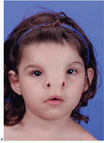
Median craniofacial hyperplasia (excess of midline tissue): This spectrum of midline anomalies includes all forms of excess tissue from a thickened or duplicated nasal septum to the more severe forms of frontonasal dysplasia (Figure 26.5).

Soft tissue midline excess may be manifested in the lip with broad philtral columns or a duplication of the labial frenulum. The nose may be bifid with a broad columella and mid-dorsal furrow. The alar and upper lateral cartilages may be displaced laterally.

Skeletal excess in a wide 0 facial cleft can be seen as a diastema between the upper central incisors. A duplicate nasal spine may exist. A keel-shaped maxillary alveolus with anterior teeth angled toward the midline creating an anterior open bite is characteristic. Central midface height is shortened. The cartilaginous and bony nasal septum is thickened or duplicated. The nasal bones and nasal process of the maxilla are broad, flattened, and displaced laterally from the midline. Ethmoidal and sphenoidal sinuses may be enlarged, contributing to symmetrical widening of the anterior cranial fossa and hypertelorism.


FIGURE 26.4. Number 0 cleft: Median craniofacial dysplasia. Patient with a “true” median cleft lip deformity.


FIGURE 26.5. Number 0 cleft: Median craniofacial hyperplasia. A-C. Patients with excessive midline tissue manifested by bifid nose and an accessory band of skin on the nasal dorsum.


FIGURE 26.5. (Continued)


Number 1 Cleft

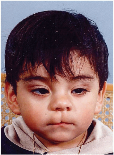
Soft tissue involvement: The number 1 cleft, similar to the common cleft lip, passes through the cupid’s bow and then the alar cartilage dome. Notching in the area of the soft triangle of the nose is a distinct feature (Figure 26.6A). The columella may be short and broad. The nasal tip and nasal septum deviate away from the cleft. When the cleft is evident medially to a malpositioned medial canthus, telecanthus may result. With accompanying cranial extension as a number 13 cleft, vertical dystopia may be present.

Skeletal involvement: An alveolar cleft would pass between the central and lateral incisors (Figure 26.6B). This paramedian cleft separates the nasal floor at the pyriform aperture just lateral to the nasal spine. The cleft may extend posteriorly as a complete cleft of the hard and soft palate. Extension of the cleft in a cephalad direction is through the junction of the nasal bone and the frontal process of the maxilla.


Number 2 Cleft

Soft tissue involvement: This other paramedian facial cleft may also begin in the region of the common cleft lip. However, the nasal deformity is in the middle third of the alar rim and distinguishes the number 2 cleft (Figure 26.7A). In the number 2 cleft the ala is hypoplastic, whereas in the number 1 cleft, the ala is merely notched at the dome. The lateral aspect of the nose is flattened and the dorsum is broad. The eyelid is not involved; the cleft passes medially to the palpebral fissure. Although the medial canthus is displaced, the lacrimal duct is usually not involved. If the cleft continues in a cephalad direction as a cranial number 12 cleft, then distortion of the medial brow is noted.

Only gold members can continue reading. Log In or Register to continue

Stay updated, free articles. Join our Telegram channel

Jun 26, 2016 | Posted by in General Surgery | Comments Off on Craniofacial Clefts and Hypertelorbitism

Full access? Get Clinical Tree

Get Clinical Tree app for offline access