Introduction
Congenital breast asymmetry can be relatively common among young women. , Although tuberous breast deformity, Poland syndrome, and structural chest wall deformities can be considered reconstructive, less extensive congenital breast asymmetry is typically corrected as a cosmetic breast procedure. This chapter will focus only on correction of congenital breast asymmetry for young women who are seeking correction of their breast asymmetry for aesthetic reasons. Often, patients will present for improvement of breast size, shape, and projection without fully realizing their current breast asymmetry, which is sometimes quite significant.
To achieve optimal symmetry and the aesthetic desires of the patient, the specifics of the asymmetry based on preoperative analysis must be identified clearly before any intervention. It cannot be understated how important it is for the plastic surgeon to fully analyze, identify, and show the patient the asymmetry and potential plans for correction. Complete correction may require a more invasive procedure with a greater scar component than the patient desires. An informed discussion with the patient will direct the procedure implemented by the plastic surgeon and the ultimate amount of needed correction.
In this chapter, the authors describe their systematic approach to address several types of congenital breast asymmetry, emphasizing preoperative evaluation, operative approaches, refinements, and secondary procedures. Correction of breast asymmetry is truly an artistic endeavor in aesthetic plastic surgery not defined by a single procedure but by using all available procedures in our armamentarium to obtain a symmetric and satisfying result for the patient.
Indications and Contraindications
In general, any congenital breast asymmetry should not be surgically addressed in the aesthetic realm until the patient is at a stable breast size and shape and thus an appropriate age. Further asymmetry and even improvement of asymmetry can be seen before completion of breast development and growth. Once breast development is stable, characteristics of the patient’s breast envelope and breast tissue may make them poor candidates for certain procedures. The same plastic surgery principles in evaluating the right procedure for a patient with symptomatic breast hyperplasia seeking a reduction, ptosis seeking a mastopexy, and hypoplasia seeking an augmentation apply to the correction of congenital breast asymmetry.
As we know, an implant of significant volume inappropriate for a specific breast footprint and a periareolar mastopexy for significant ptosis will lead to the same poor outcomes as doing these procedures for their primary purposes. Another relative contraindication could be the patient wishing for correction of breast asymmetry who had baseline prepartum asymmetry that has worsened during and after her pregnancy and plans for further pregnancies. Even after appropriate correction, these patients may have a recurrence after further pregnancy and this needs to be fully discussed before any intervention.
Preoperative Evaluation and Special Considerations
In the senior author’s practice, a total of four categories for less extensive breast asymmetry have been classified and thus any treatment recommendations could be based on this classification. We have found that the classification is quite useful and can be used to select appropriate procedures for each patient.
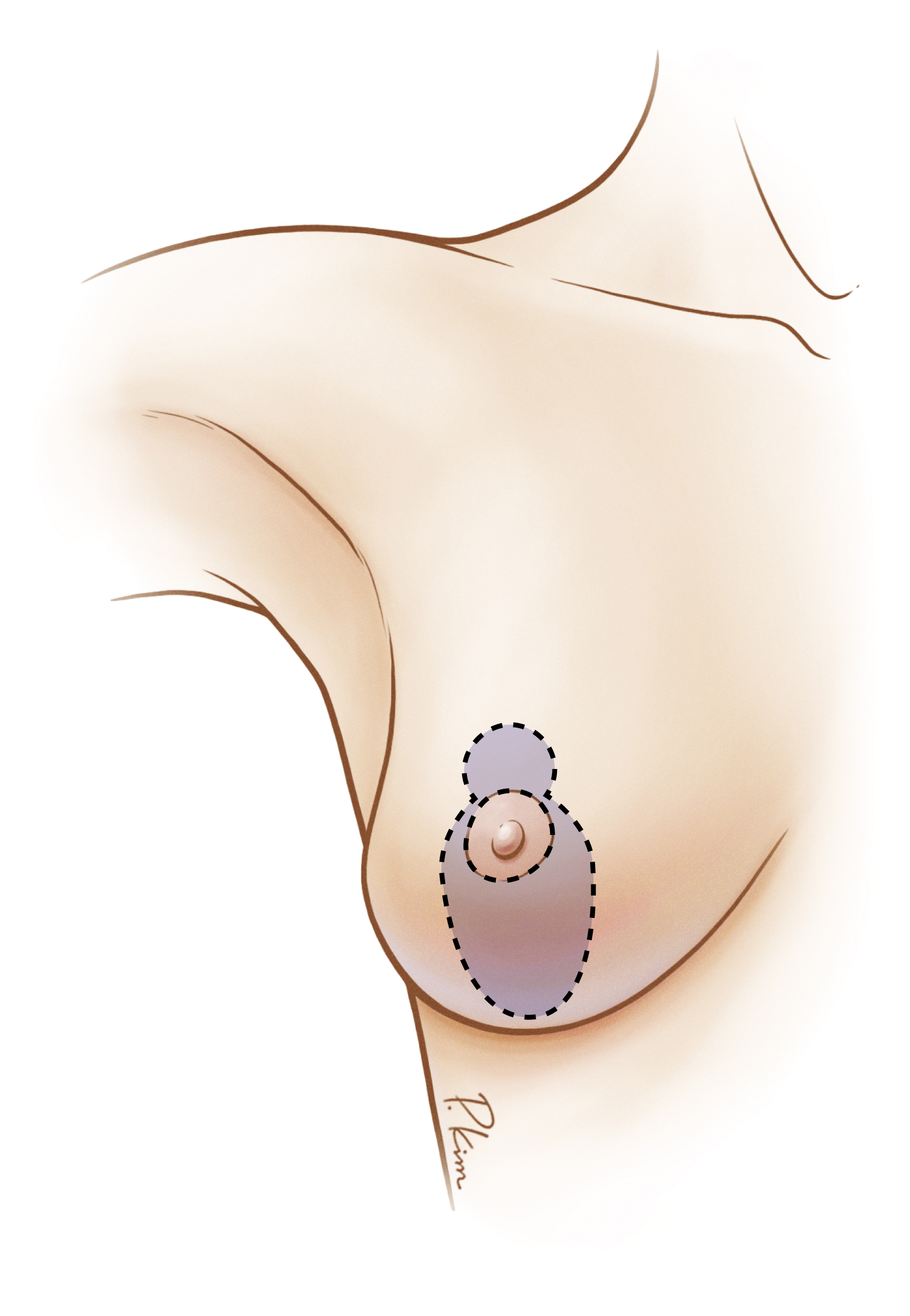
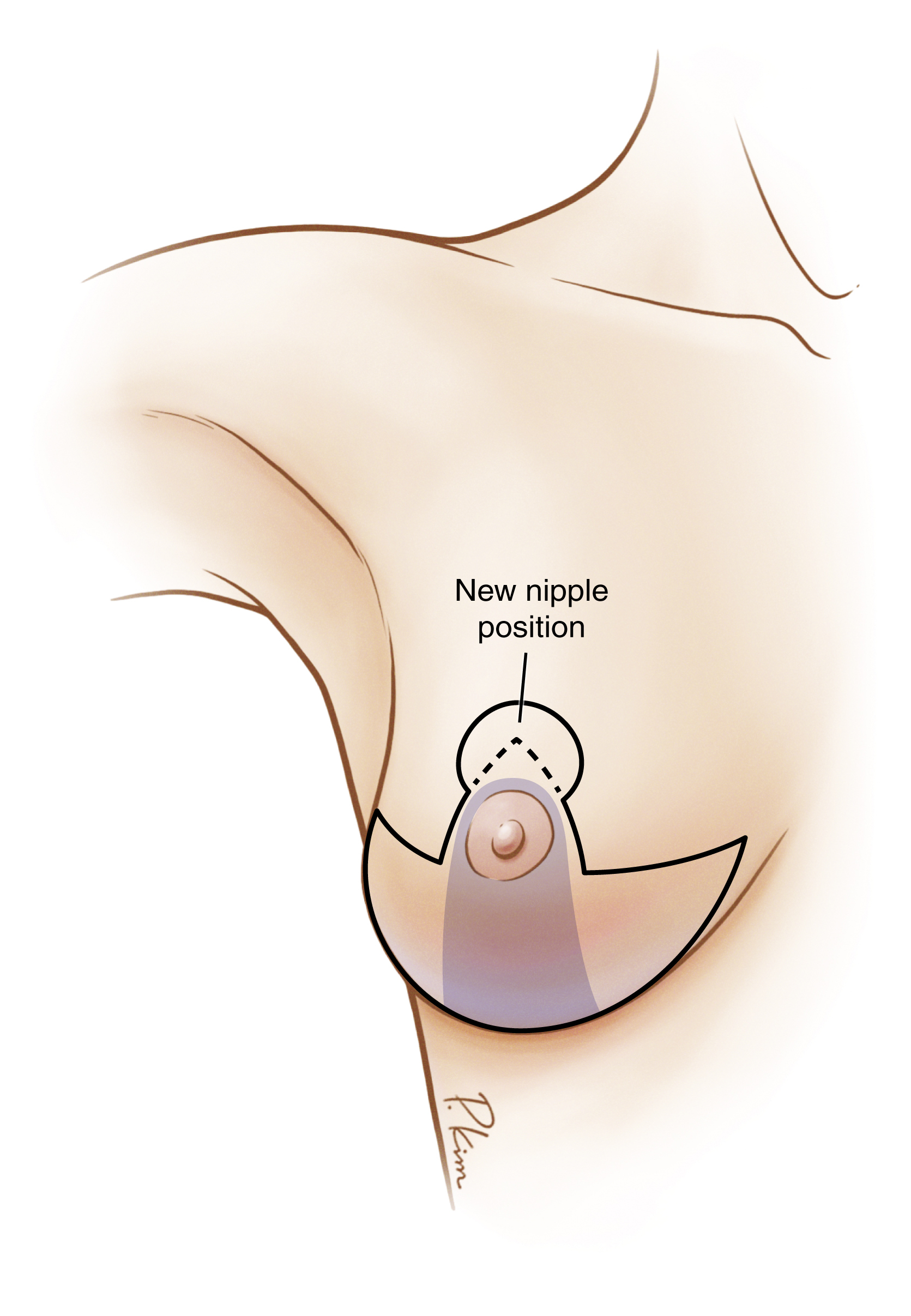
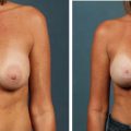
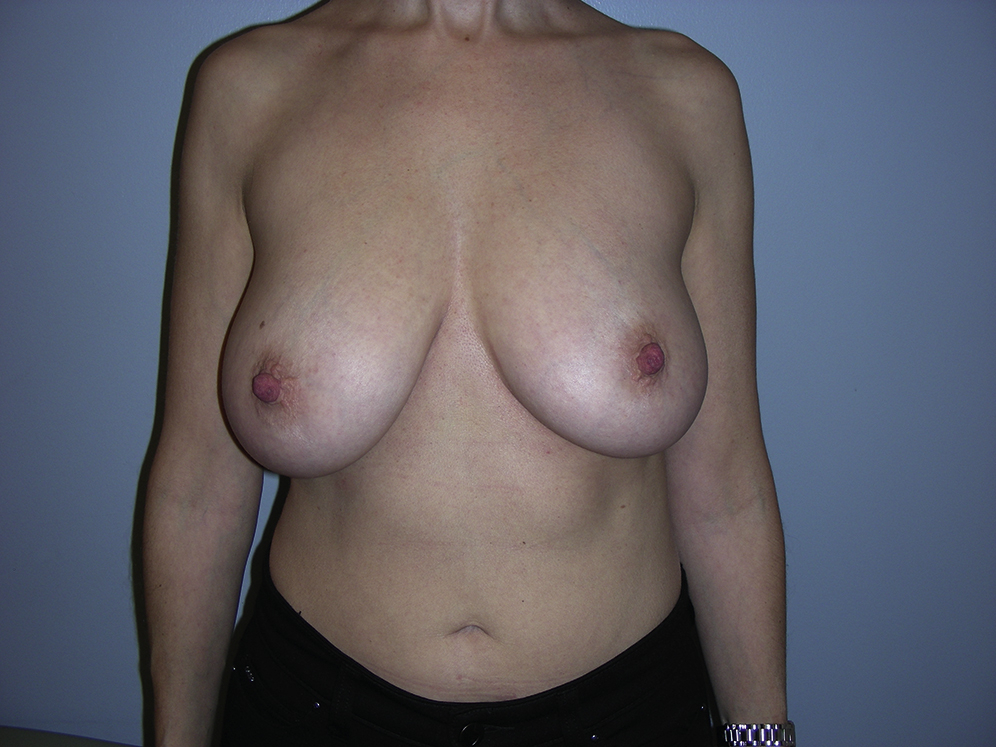
Type 1 is bilateral asymmetric breast ptosis. The ptosis may be classified as the same or different Regnault grade, but the different degrees of ptosis are clear to the plastic surgeon and all discrepancies should be pointed out to the patient. It should be made evident to the patient that different procedures and hence scar patterns could be used on each side to achieve a symmetric result in regard to breast shape ( Fig. 24.1 ).
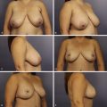
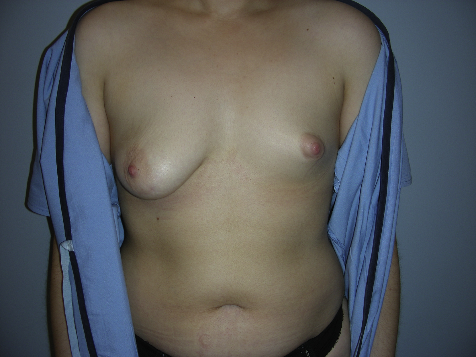
Type 2 is bilateral breast hypoplasia of different sizes. An attempt to classify the approximate amount of hypoplasia on each side quantitatively is recommended, to have an informed discussion with the patient that correction of asymmetry will likely require different sizes of implants and possibly even different projected implants. Despite getting an estimate of size discrepancy quantitatively, commitment to a specific size and projection preoperatively is not recommended ( Fig. 24.2 ).
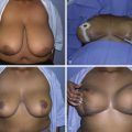
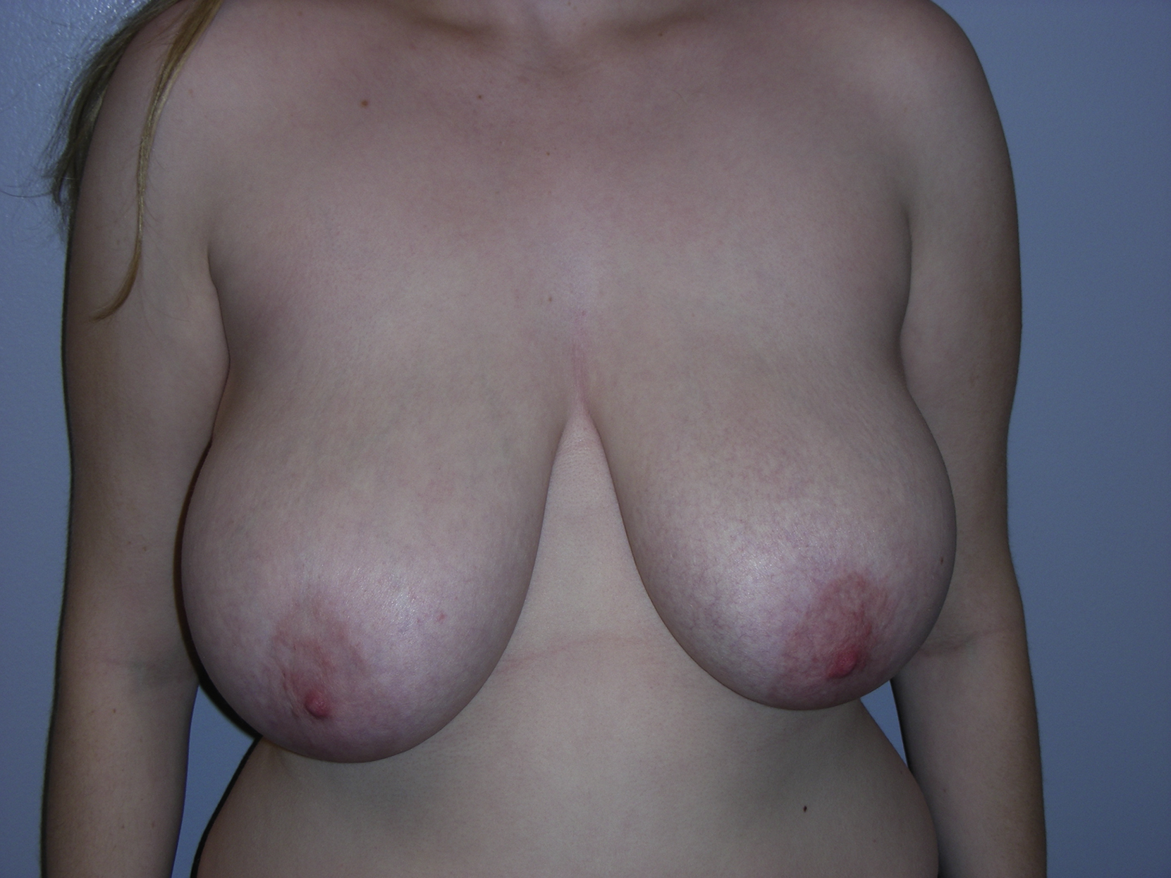
Type 3 is bilateral breast hyperplasia of different sizes. These patients are not uncommon in the plastic surgery practice and often present with concerns of symptomatic macromastia, although unaware of their breast asymmetry. It is critical to identify these size discrepancies and inform the patient. As with type 1, it should be made evident to the patient that different procedures and hence scar patterns could be used on each side to achieve a symmetric result ( Fig. 24.3 ).
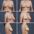
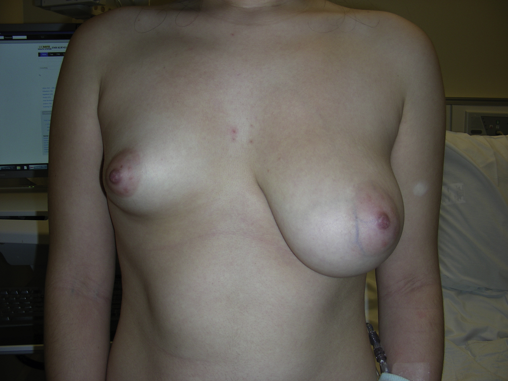
Type 4 is typically the most difficult to correct surgically; these patients have one hyperplastic breast and one hypoplastic breast. For those cases, the patient will require a reduction/lift technique on one breast and an augmentation/lift technique on the other breast to obtain symmetry. These cases require the most formal operative planning and discussion with the patient that the good end result can be time-consuming and difficult to obtain ( Fig. 24.4 ).
Not a separate type but equally important in its own right is differences in inframammary fold (IMF) height. Discussion and correction of discrepancies in IMF height is important for all the asymmetry types.
Surgical Techniques
Relevant Surgical Anatomy
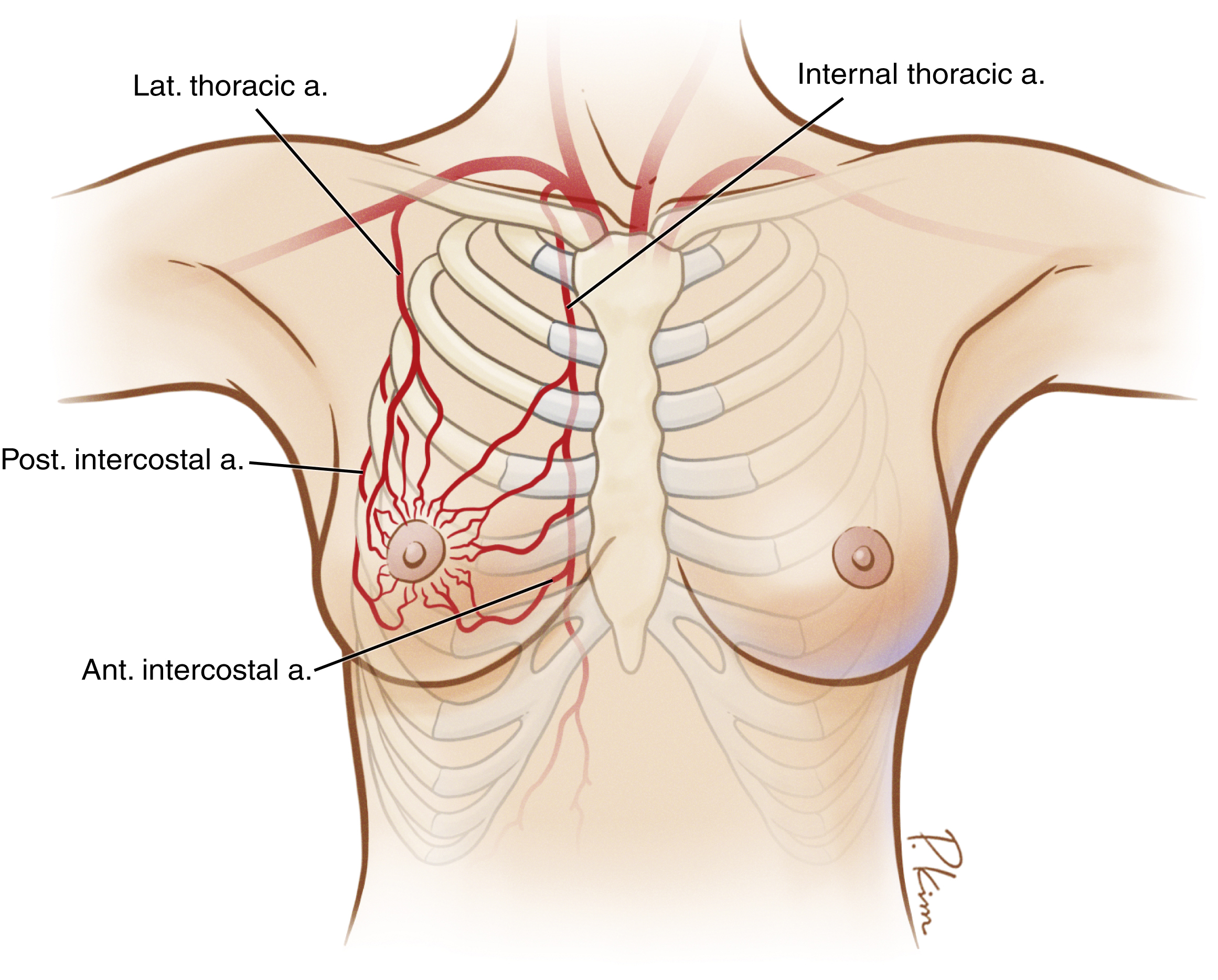
The breast receives its blood supply from several directions. However, the main blood supply to the breast is based on medial branches of the internal mammary artery ( Fig. 24.5 ). The superomedial perforators from the internal mammary vessels are particularly robust and account for some 60% of the total breast blood supply. This rich blood supply allows for designs of various mastopexy or reduction techniques, ensuring the viability of the nipple–areola complex or skin flaps after surgery. The veins of the breast rarely accompany the arteries. Much of the breast is drained by a superficial venous system that lies just under the dermis. The nipple is primarily innervated by the medial and lateral branches of the fourth intercostal nerve. However, the third and fifth intercostal nerves contribute as well.
Common Techniques
Several common breast surgical procedures are used in the senior author’s practice for correction of congenital breast asymmetry. These are breast augmentation with implant by inframammary approach, mastopexy (vertical or inverted-T or periareolar pattern), and breast reduction (medial pedicle or inferior pedicle technique). In addition, a combination of breast augmentation and mastopexy also has been commonly performed for the same purpose.
To optimize outcomes, we prefer to work on both sides simultaneously to achieve symmetry because each asymmetric breast is commonly not in the “normal” position as well. This allows the surgeon to have more freedom and make any necessary refinements to obtain the greatest possible symmetry after the major portion of the procedures is completed.
Type 1 Breast Asymmetry
When addressing type 1 asymmetric breasts with differing degrees of bilateral ptosis, the surgical technique implemented on the more ptotic side is typically a vertical or inverted-T mastopexy or even breast reduction, as indicated ( Figs. 24.6 and 24.7 ). The chosen procedure will be based on the degree of ptosis and what the surgeon and patient agreed to proceed with during the preoperative evaluation. A vertical or periareolar mastopexy is often executed on the less ptotic side ( Fig. 24.8 ), but again this is guided by degree of ptosis and preoperative discussion with the patient. The skin pattern of mastopexy for each side can be determined intraoperatively by the tailor-tacking technique. Once the appropriate procedure for the degree of ptosis is implemented, with the patient in the sitting position, further refinements are made based on the nipple position and breast shape using the same principles described for mastopexy.