and Peter M. Prendergast2
(1)
Elysium Aesthetics, Bogota, Colombia
(2)
Venus Medical, Dublin, Ireland
Introduction
Effective high-definition body sculpting employs advanced techniques and extensive superficial lipoplasty and should not be attempted by beginners. If the procedure is not planned and performed with great attention to detail and with proper postoperative care, the complication rate will be higher. The patient should be made aware during informed consent that high-definition lipoplasty is different to conventional lipoplasty. During the procedure, controlled irregularities are deliberately created to enhance definition. Patients who may not be motivated to attend frequent postoperative visits should be excluded since postoperative therapies play an important role in optimizing results and minimizing complications. The patient must also understand the importance of constant compression with foam vests and garments in the postoperative period. Most of the complications associated with high-definition body sculpting can also arise following conventional lipoplasty techniques. They are described in this chapter. The use of third-generation ultrasound-assisted lipoplasty (VASER) technology is important for successful high-definition lipoplasty and may also reduce excessive blood loss, prolonged edema, as well as stimulate skin retraction [1, 2].
General Liposuction Complications
The following general complications are not specific to high-definition lipoplasty and may arise following any form of surgical fat removal.
Bleeding
Excessive bleeding during or following high-definition body sculpting is rare. Using the tumescent technique, saline causes profound vasoconstriction so the emulsification and removal of fat is occurring in an almost bloodless field. Although a creamy, bloodless aspirate is typical during tumescent VASER lipoplasty, a bloody aspirate may be produced in very fibrous areas such as the back, upper abdomen, or male breasts. If the aspirate is excessively bloody, the area should be avoided or more tumescent fluid should be infiltrated.
To exclude patients who potentially have a bleeding abnormality, the preoperative investigations should include at least a full blood count including platelet count, liver function tests, prothrombin time (PT), and activated partial thromboplastin time (APTT). Any abnormality should be repeated and the patient should be referred to a hematologist for further investigation. A positive family history of bleeding diathesis also warrants detailed coagulopathy studies. Preoperatively, patients should not take any medications, vitamins, herbals, or supplements with antiplatelet effects for at least 7–10 days. These include aspirin, nonsteroidal anti-inflammatory drugs, vitamin E, ginseng, ginger, ginkgo biloba, and garlic.
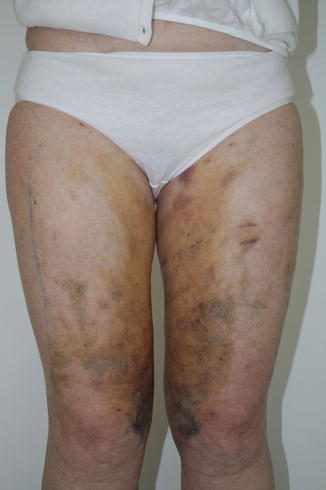
Sequelae of intraoperative and postoperative bleeding depend on the volume of blood loss, condition of the patient, treatment area, and presence of drains and compression. Open or closed drains serve to reduce the likelihood of hematoma by evacuating blood. The application of compression garments is essential immediately postoperatively in order to facilitate hemostasis and compress small vessels. Although some ecchymosis is normal, extensive ecchymosis may occur in areas more prone to have bruising, particularly in the thighs (Fig. 20.1). Resolution of ecchymosis occurs without intervention, although oral or topical arnica Montana may accelerate healing [3]. A small hematoma may resolve without intervention. A large hematoma can be evacuated or aspirated once it liquefies. Untreated, a large hematoma will become organized and form a mass, seroma, and chronic pseudocyst requiring aspiration [4].


Fig. 20.1
Extensive bruising over the thighs following ultrasound-assisted lipoplasty
Infection
Infection is unusual following lipoplasty and high-definition body sculpting. Sterile technique using properly sterilized instruments in appropriately selected patients serves to keep the incidence of infection very low. The components of tumescent fluid (lidocaine, epinephrine, and sodium bicarbonate) also have antimicrobial effects on bacteria, mycobacteria, and fungi [5]. Despite this, serious, life-threatening infections following liposuction have been reported [6, 7].
Patients who are immunocompromised will have an increased risk of infection and should either be excluded from treatment or their condition controlled and closely monitored in the perioperative period. Preoperative investigations to identify these pathologies like diabetes or human immunodeficiency virus (HIV) include fasting glucose, testing for HIV I and HIV II, and hepatitis C. Patients taking systemic corticosteroids are contraindicated from high-definition body sculpting, since the risks of impaired healing and infection outweigh the benefits. Smokers should be advised not to smoke for at least 1 month before and for 1 month after surgery. Prophylactic antibiotics, such as a cephalosporin, should commence before surgery and be continued for 5 days. An alternative can be used for patients allergic to penicillin. Some surgeons prefer intravenous antibiotics at the time of surgery.
A superficial infection around the incision site following high-definition body sculpting will manifest as blanching erythema, heat, and tenderness. The offending organism is usually staphylococcus or streptococcus. Fluid, exudate, or pus should be sent for culture and sensitivity in order to ensure appropriate antibiotic coverage. A swollen, red, mass that appears weeks or even months after surgery should raise the suspicion of an atypical mycobacterial infection. These masses may require drainage or excision and prolonged antibiotic therapy. Necrotizing fasciitis represents a severe life-threatening streptococcal group A infection or mixed bacterial infection that leads to thrombosis of the subcutaneous vessels and spreading gangrene. Timely treatment with antibiotics, surgical debridement of necrotic tissue, and hyperbaric oxygen therapy may prove curative.
Necrosis
Necrosis of tissue and skin may occur following aggressive superficial lipoplasty if the subdermal vascular plexus is compromised. This is more likely if the patient is a smoker, in secondary cases, or if sharp cannulae are used. If the ultrasonic probe is not continuously moving in the tissues, or if it is activated in dry tissues, mechanical energy will be converted to heat and a burn may result. In order to minimize the incidence of necrosis, very superficial maneuvers such as pinching or compression should be limited to areas where shadows are required such as over the linea alba or linea semilunaris and not performed extensively over the abdomen. Additionally, a burn or necrosis is rare when pulsed ultrasonic delivery to the superficial tissues is used using a gentle dynamic technique in wet tissues. The postoperative compression foam and garment should be tailored to fit the patient properly to avoid excessive compression that may compromise perfusion to the skin and superficial tissues.
Seroma
A seroma is an abnormal collection of fluid in the subcutaneous tissues that occurs as a result of trauma, burns, or friction following lipoplasty. It is usually an inflammatory exudate, but may also be comprised of lymph [8]. An untreated hematoma may also become a seroma over time. Seromas are more common in overweight or obese patients with large abdomens. Large seromas are uncommon following high-definition body sculpting because the patients are usually not obese [9]. Diagnosis is either by clinical examination or ultrasound examination. Early seromas can be drained by needle aspiration followed by compression. Drainage may be required every few days until it resolves. A chronic seroma that persists longer than 1 month may require aspiration and injection of room air into the cavity, curettage, or formal excision. Ultrasound-guided drainage is more accurate than blind aspiration [10].
Thromboembolism
Risk factors for deep venous thrombosis (DVT) and pulmonary embolism (PE) should be identified during the consultation and assessment. These include age over 40 years, obesity, previous history of thromboembolism, cancer, smoking, and estrogen therapy. Prolonged surgery and postoperative immobilization also increase the risk. For lengthy procedures performed under intravenous sedation or general anesthesia, perioperative antithrombotic prophylaxis will reduce the risk of DVT and PE. In general, if the procedure is performed purely under tumescent local anesthesia where the patient is moving during the procedure, prophylaxis is not required. Patients should mobilize as soon as possible postoperatively and stay well hydrated. Measures to reduce the risk of thromboembolism associated with high-definition body sculpting include discontinuing estrogens for 3 weeks before and after surgery, ensuring the patient wears thromboembolic deterrent (TED) or antiembolic/antithrombotic stockings, subcutaneous low molecular weight heparin toward the end of the procedure and postoperatively for patients undergoing procedures estimated to last longer than 1 h under sedation or general anesthesia, and excluding high-risk patients from treatment. A DVT may manifest as calf swelling, tenderness, or pain on passive dorsiflexion of the foot. A duplex scan should be performed to assess the deep venous system and make the diagnosis. Symptoms and signs of PE include tachycardia, dyspnea, tachypnea, and pleuritic chest pain. Several cases of fatalities due to pulmonary embolism associated with liposuction have been reported [11, 12]. If suspected, therapy may be commenced even before a definitive diagnosis is made with imaging such as ventilation-perfusion lung scan and CT pulmonary angiography.
Pulmonary Edema
The preoperative history and examination should identify patients with preexisting cardiac disease. An electrocardiogram, chest X-ray, and echocardiogram can be used to investigate cardiac function. Patients with cardiac failure should not undergo tumescent lipoplasty due to the large volumes of fluid infiltrated during the procedure. Even in healthy patients, injudicious use of intravenous fluids as well as large volumes of subcutaneously infiltrated fluid can cause fluid overload and pulmonary edema [13]. Fluid balance should be monitored carefully during high-definition body contouring where large surface areas are being treated and large volumes of tumescent fluid are required. Usually intravenous fluids are not required except to maintain a patent intravenous line.
Lidocaine Toxicity
Lidocaine can be administered safely in tumescent anesthesia in doses up to 55 mg/kg [14]. Reducing the maximum dose to 45 mg/kg is prudent in order to reduce the incidence of lidocaine toxicity. Toxicity occurs when lidocaine is absorbed systemically and appears as perioral tingling, numbness of the tongue, dizziness, nausea, and vomiting. With increasing plasma levels, cardiac toxicity can occur. The treatment of lidocaine toxicity is supportive. It is important to remember that since lidocaine is absorbed very slowly from the fat compartment, peak plasma concentrations occur only 12–18 h after administration. Drugs that are metabolized through the cytochrome P450-3A4 enzyme pathway compete with lidocaine and have the potential to increase toxicity [15]. These medications should be discontinued for at least 2 weeks before tumescent anesthesia. During tumescent anesthesia, the rapid infiltration of fluid may increase serum levels of lidocaine since it takes 12–15 min for epinephrine to cause full vasoconstriction. A lidocaine concentration of 0.05–0.1 % is safe and effective for most areas and indications. For high-definition body sculpting under local anesthesia, a concentration of 0.1 % (1,000 mg lidocaine in each 1 L bag physiologic saline) is often required in the superficial tissues to ensure full anesthesia during detailed sculpting. If the procedure is performed under general anesthesia, the lidocaine dose is reduced to about 25 % or even less. Lidocaine administered to patients under general anesthesia provides postoperative analgesia for several hours due to the lipophilic nature of the lidocaine molecule.
Perforation
Penetration of the probe or cannula through the abdominal wall can occur during any lipoplasty procedure on the abdomen, particularly if the procedure is performed under intravenous sedation or general anesthesia. Several cases of intestinal perforation following abdominal lipoplasty have been reported [18–20]. Intestinal perforation causes peritonitis and may lead to septic shock, necrotizing fasciitis, and death. A perforation can be the result of the cannula passing through a defect in the abdominal wall, into an undiagnosed abdominal wall hernia, or careless movement of the cannula in a vertical, rather than horizontal, direction. An ultrasound scan of the anterior abdominal wall should be performed preoperatively if an hernia is suspected or if a scar from previous surgery appears tethered or depressed [18]. The peritoneum could be adherent to the skin in such cases. Intraoperatively, the nondominant hand should always feel the tip of the probe or cannula as gentle strokes are made through the tissues. Particular care should be taken near the costal margin to avoid penetration of the thoracic cavity. Treating the upper abdomen using access incisions in the inframammary crease ensures the cannula is oriented inferiorly over the costal margin, reducing the risk of inadvertent intrathoracic penetration. Care should also be taken to protect the posterior costal margin when treating the flanks or back. As well as perforation of a viscus, vessels may be injured causing intra-abdominal or retroperitoneal bleeding [21


Stay updated, free articles. Join our Telegram channel

Full access? Get Clinical Tree


