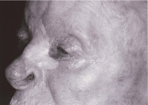
5 The use of local and regional flap reconstruction in the repair of facial defects is a mainstay of facial plastic and reconstructive surgery. The surgeon has various options available when assessing treatment of a soft-tissue defect. Each must be considered in the context of multiple factors, such as knowledge of the anatomy, vascular supply, and tissue quality available. The surgeon is required to select a course of action tailored to the individual that may include healing by secondary intent, primary closure, skin grafts, local and regional flaps, distant flaps, and free tissue transfer. Each patient should be assessed in terms of age, race, gender, occupation, nutrition, prior irradiation, skin type, habits, and general health. Cosmetic success and survival of a local or regional flap depend on considering all these variables. The appropriate surgical approach must be based on both the distinctive characteristics of the defect and the multiple unique factors presented by each patient. The surgeon should strive to satisfy the patient’s reasonable expectations and goals, understanding that these goals may differ greatly from patient to patient, based on the individual’s priorities, age, gender, and health. No precise statistics regarding complication rates are available, but adverse outcomes involving locoregional flaps can best be avoided by meticulous preoperative planning, a frank discussion with the patient about the relative risks and benefits associated with each reconstructive approach, and sound technique. Before proceeding with treatment, the surgeon must fully evaluate the dynamic spectrum of economic, psychosocial, and general health issues, coupled with the reconstructive dilemma each patient presents. The simplest approach to repair is not always the best choice for the patient, as sometimes a more complex approach will result in a better cosmetic and functional result. Sometimes primary closure is simple and effective. Healing by secondary intent is an excellent option for small defects involving concave skin surfaces, such as the medial canthus, temple, and occasionally the nasal alar groove.1 Yet healing by secondary intent necessitates a long period of wound care, which some patients may not find acceptable. Conversely, wounds involving convex skin surfaces tend to heal poorly by secondary intent and are therefore more complex in their management. Locoregional flaps typically provide excellent skin color and texture match, and they may be the best option when primary closure and secondary intent closure are not the best options. Reconstruction using locoregional flaps can commonly be performed in a single stage, and the donor sites can be closed primarily with little morbidity. Locoregional flap complications can stem from poor preoperative planning, infection, hematoma, or patient-specific factors that compromise the vascularity of the tissue or the healing of the scars. The choice of an appropriate flap to close a specific defect is complex and is not the point of this chapter. One is referred to other textbooks on reconstruction for discussion of flap choices. Yet, one must understand that surgery starts with the preoperative planning. Incisions have to be planned parallel to relaxed skin tension lines, in aesthetic unit and subunit boundaries, or in the midline of the face. An undersized flap can result in distortion of critical anatomic structures, causing ectropion, alar retraction, or retraction of other surrounding structures. Deep wounds must be reconstructed with both adequate structural support to overcome scar contracture and adequate bulk to fill the defect, but not so much bulk as to cause a contour deformity. Deep layer closure is critical to decrease the dead space where blood or scar tissue can accumulate, as well as to decrease the tension on the skin closure so that wounds heal with optimal scars. Although most complications can be avoided, others are simply the result of indiscriminate odds. This chapter will focus on the complications of locoregional flaps, ways to decrease the likelihood of a complication, and management in the event of a complication. The term flap was derived from the Dutch word flappe in the 16th century, meaning something that hung broad and loose, secured by only one side. The first historical mention of flap surgery dates to a more remote time, around 600 BC, when Sushruta Samhita, believed to be part of one of the four Vedas, described what he had learned from his mentor Dhanwantri. He eloquently described using a cheek flap for nasal reconstruction. The traditional Indian technique for rhinoplasty using a flap from the adjoining forehead can be traced back to approximately 1440 AD. Later surgical methods using flaps to repair defects progressed in distinct periods. During the First and Second World Wars, pedicled flaps were used extensively. Subsequently the procedure of axial pattern flaps (flaps with a named blood supply) was developed in the 1950s. In the 1970s, the distinction between axial and random flaps (unnamed blood supply) was first recognized. The ensuing advances include the use of cutaneous, fascio-cutaneous, muscle, musculocutaneous, and osseous tissue types in flap surgeries. The defining characteristic of a skin flap is that its survival in the recipient bed is predicated upon a functioning intravascular circulation. Historically, the design of skin flaps was governed by a set of length-to-width ratios (5:1 for the face due to its abundant blood supply). Local flaps can easily be advanced, pivoted or interpolated, thereby allowing a greater length-to-width ratio. However, in the 1970s, Daniel showed that increasing the width of a flap did not increase the surviving length.2,3 Rather, the amount of blood supply incorporated into a flap’s width dictates the flap’s surviving length. Daniel and Williams defined the blood supply to the skin as being from two types of arteries: (1) musculocutaneous arteries or (2) direct cutaneous arteries (now further subdivided4). The skin is supplied by two vascular plexuses. The deep vascular plexus, or subdermal plexus, is found at the junction of the dermis and subcutaneous fat. The superficial vascular plexus courses in the superficial aspect of the reticular dermis, giving off capillary loops within the dermal papillae. Musculocutaneous arteries are branches of segmental vessels arising from the aorta that supply blood to muscle and continue through the underlying muscle to the overlying dermal plexi. For the majority of their course, musculocutaneous arteries run perpendicular to the skin, supplying only small areas of the skin. Direct cutaneous arteries are branches of segmental or muscular arteries that pass through fascial septae between muscles to supply the enveloping fascia and overlying skin. Arteries run parallel to the skin, supplying a large area of the skin.5 Random flaps (e.g., bilobed, note, z-plasty) are the most commonly used in local flap repair of facial defects. The vascular supply to random flaps, which consist of skin and subcutaneous fat, arises mainly from multiple musculocutaneous arteries from the subdermal plexus at the flap’s anatomic base. Therefore the appropriate plane of dissection is at the level of the subcutaneous fat. Axial flaps derive their blood supply from named direct cutaneous arteries. Therefore, axial flaps allow for greater length and reliability, with less concern for width or a delay procedure.6 Surgically, the plane of dissection is deeper to incorporate fascia and must align along a distinct septocutaneous vessel. Examples of axial flaps include the paramedian forehead flap, based on the supratrochlear artery, and the nasiolabial flap, based on the angular artery. Reconstruction of facial soft-tissue defects requires the surgeon to have knowledge of wound healing physiology, soft-tissue handling, and head and neck reconstructive surgical techniques. Poorly designed flaps can lead to an array of complications, ranging from minor cosmetic deformities to functionally debilitating abnormalities (Fig. 5.1). The planning of surgical incisions should adhere to the elementary principles of relaxed skin tension lines (RSTL) and lines of maximal extensibility (LME), which typically lie perpendicular to each other. Incisions heal better when placed parallel to or in the RSTLs, and wound tension is minimized if donor tissue is transferred along LMEs.7,8 Additionally, wounds placed in aesthetic unit and subunit boundaries, along hairlines, and in the midline of the face heal with less perceptible scars. Wounds aligned with RSTLs are not subjected to repetitive tension and therefore reduce the risk of scar hypertrophy.9,10 Fig. 5.1 Patient who demonstrates multiple late sequelae of poor defect repair choices, including alar retraction, ectropion, and color mismatch between the skin graft and surrounding tissue. Wound immobilization during healing will result in better cosmetic outcomes. Two techniques commonly employed to minimize wound tension during healing are undermining and using deep sutures. Undermining, however, is not always the best method to relieve excessive tension. It has been shown that beyond 4 cm, undermining has no effect on tension. Furthermore, animal studies suggest there is a correlation between excessive skin undermining and an increased incidence of flap necrosis.11 Deep sutures help to relieve tension and make wound edge eversion easier. Wound edges that are everted at the time of closure result in better scars. Recently, Gassner et al. have reported statistically significant results in improved scar appearance following use of botulinum toxin injections at the time of surgical repair.12–15 Botulinum toxin decreases the tension on a wound by temporarily chemodenervating the muscles pulling on the wound. Gassner and Sherris recommend injecting the surrounding muscles at the time of wound closure to produce an improved scar. This theory has been proven in a blind, placebo-controlled trial in animals and humans with forehead defects and in an open trial with perioral wounds. Further studies using this innovative technique are underway and are promising. Careful attention to the aforementioned principles of minimal wound tension is vital when the surgeon is designing flaps for defects involving the nasal ala, the eyelids, and the vermilion border of the mouth. These areas are extremely sensitive to unusual skin tension.16–22 Increased tension on the nasal ala from a poorly designed flap may result in alar retraction, nasal obstruction, or both (Fig. 5.1). Poor flap design in the eyelid region may lead to ectropion, epiphora, or lagophthalmos, which may further result in exposure keratitis of the cornea with possible visual impairment. If the vermilion border of the lip in the perioral region is subjected to excessive tension, oral competence and speech may be compromised. These defects, aside from leading to a functional impairment, are very difficult to revise. Therefore, it is crucial to examine these anatomic structures meticulously after insetting of the flap intraoperatively. Increased tension should be relieved, even if this requires using an alternate method of closure. Consideration of placing structural grafts under the eyelid or nasal flaps to overcome the wound contracture is an effective measure to prevent contracture of these important anatomic structures. General wound-handling principles for soft tissue should be meticulously observed during surgery of the face. Tissue should be handled with skin hooks and fine toothed forceps to minimize blunt trauma.7,8 Tissue layers need to be properly sutured to eliminate dead space. Meticulous hemostasis should be obtained with bipolar electrocautery. Overly aggressive electrocautery, however, can increase the risk of flap necrosis. Furthermore, excessive thinning of the donor tissue reduces flap viability. Minimizing wound tension will avert damage to axial vessels and decrease the occurrence of traumatic dissection. The design of a facial flap must always assimilate the knowledge of the underlying anatomy to avoid injury to vulnerable anatomic structures. Knowledge of the locations of the major vessels and nerves of the face, as well as the tissue layers in which they reside, is essential when considering flap elevation in these regions. Invariably, small sensory nerves will be transected at the margins of a flap, but the mild loss of cutaneous sensation is not usually clinically significant. However, the great auricular nerve, which is located anterior and superficial to the sternocleidomastoid muscle, can easily be injured unless one stays superficial to the fascia of the sternocleidomastoid muscle. If the nerve is injured, hypesthesia or paresthesia of the affected ear may occur. The infraorbital nerve, which supplies sensation to a large area of the midface, is also susceptible to injury, especially in eyelid and nasolabial flap reconstruction. A far more serious injury can occur if the facial nerve is injured. The temporal branch of the facial nerve is very close to the surface as it crosses the zygomatic arch in the temporal region.23–25 It is vulnerable to injury in this area, especially in elderly patients with thin skin. Transection of the temporal branch of the facial nerve can result in the loss of forehead furrows and lead to eyebrow ptosis. Injury to branches supplying the orbicularis oculi muscle may result in difficulty closing the affected eye. The zygomatic branch of the facial nerve is less vulnerable as it crosses the zygoma, because it does so in a deep plane. Transection will result in eyelid ptosis and ectropion. Injury to the buccal branch of the facial nerve along its course from the parotid gland to the orbicularis oris muscle may result in facial asymmetry, an inability to pucker the lips, or synkinesis. The marginal mandibular nerve is most vulnerable at the angle of the jaw as it courses from under the parotid gland. Facial nerve damage can result in both cosmetic deformity and significant functional impairment, particularly around the corner of the mouth. To avoid these devastating consequences of facial nerve injury, flaps in these regions should be elevated and undermined in a subcutaneous plane to avoid disruption of the superficial musculoaponeurotic system (SMAS) or platysma muscle. Generally, the blood supply to the skin far exceeds the skin’s nutritional demands. Blood flow to the skin is integral for thermoregulatory function and therefore fluctuates widely as demand varies. This oversupply allows for random flap viability. The face, in particular, has a rich blood supply from the subdermal plexus. Therefore, local random flaps on the face survive with fewer complications due to vascular compromise than local flaps in other regions of the body. As previously mentioned, axial flaps in the face are based on the superficial temporal, supraorbital, supratrochlear, transverse facial, and facial angular arteries, which ensure a robust vascular supply, so axial flaps can therefore be designed with a longer flap length than random flaps.26–32 Probably the most common complication of locoregional flap reconstruction is postoperative ischemia. Ischemia, by definition, is a compromise of the vascular supply that is inadequate to provide sufficient tissue oxygenation and results in lessened flap viability. Flap ischemia may be caused by multiple factors, including poor flap design, infection, venous congestion, excess tension, or underlying vascular disorders.33,34 Ischemia may lead to tissue compromise due to hypoxia, complicated by dehiscence, infection, free radical production, and ultimately flap necrosis.35–39 Smoking greatly increases the risk of flap ischemia. Thus, patients are encouraged to stop smoking two weeks before and two weeks after flap surgery. Active bleeding at a flap’s distal margin during surgery is typically a good sign of tissue perfusion.40–42 However, perfusion cannot be correlated with adequate tissue oxygenation.43 Tissue requirements and hemoglobin saturation change in response to trauma, the presence of free radicals, acidosis, and thromboxane A2.44–46
Complications Involving Locoregional Flap Reconstruction of Facial Defects
 Brief History
 Vascular Anatomy
 Preoperative Planning
 Complications and Their Management
Ischemia
![]()
Stay updated, free articles. Join our Telegram channel

Full access? Get Clinical Tree


