This article reviews common complications encountered in the setting of facial trauma. Many complications are the result of the primary injury, and a facial plastic surgeon should be able to quickly identify these to prevent further morbidity. Common pitfalls and controversial topics are presented, as well as an overview of treatment for many complications.
Key points
- •
Intracranial and ocular injuries are common with severe facial fractures, and must be quickly identified and appropriately treated.
- •
Meticulous fracture reduction and implant placement are paramount in preventing postoperative complications.
- •
Complications of rigid fixation are typically due to fixation of inadequately reduced fractures.
- •
Close postoperative assessment allows for early recognition of complications, and provides the opportunity to intervene when necessary to achieve better long-term outcomes.
Introduction
Complications are common in the facial trauma setting, and there are several causes. All facial trauma surgeons should be knowledgable about potential associated intracranial and ocular injuries and how to prevent further morbidity. A multidisciplinary approach is often required, and early consultation with appropriate specialists is recommended. An understanding of common posttraumatic complications will guide surgical management. The most common complications of facial trauma are summarized in Tables 1 and 2 .
| Early | Late/Postoperative | |
|---|---|---|
| Soft tissue | Infection/abscess | Scar contracture |
| Loss of soft tissue | Facial deformity | |
| Unfavorable scarring | Infection/abscess | |
| Brain | Dural laceration | |
| Cerebrospinal fluid (CSF) leak | ||
| Hematoma (epidural, subdural, subarachnoid, intracerebral, intraventricular) | ||
| Diffuse axonal injury | Recurrent CSF leak | |
| Edema | Meningitis | |
| Traumatic brain injury | Brain abscess | |
| Edema | ||
| Concussion | ||
| Foreign body | ||
| Nasolacrimal apparatus | Lacrimal injury | Epiphora |
| Dacrocystitis | ||
| Parotid gland | Hematoma | Sialocele |
| Infection | Salivary fistula | |
| Sialocele | Parotitis | |
| Salivary fistula | Chronic pain | |
| Abscess | Frey syndrome | |
| Facial deformity | ||
| Eye | Traumatic optic neuropathy | Persistent diplopia |
| Retrobulbar hematoma | Enophthalmos | |
| Globe rupture | Exopthalmos | |
| Vision loss | Lower-lid malposition | |
| Diplopia | Exposure keratitis | |
| Muscle entrapment | Blindness | |
| Enophthalmos | Sympathetic ophthalmia | |
| Corneal abrasion | ||
| Superior orbital fissure syndrome | ||
| Orbital emphysema | ||
| Oculocardiac reflex (bradycardia) | ||
| Blindness | ||
| Sympathetic ophthalmia | ||
| Bone | Delayed union | |
| Fracture | Nonunion | |
| Bone loss | Malunion | |
| Infection/osteomyelitis | ||
| Dentition | Malocclusion | Malocclusion |
| Direct injury to tooth root | Tooth loss | |
| Avulsion | Infection/abscess |
| Early | Late/Postoperative | |
|---|---|---|
| Skull fracture | Traumatic brain injury | Recurrent CSF |
| Meningitis/brain abscess | Anosmia | |
| CSF leak | Meningitis/brain abscess | |
| Pneumocephalus | Seizure | |
| Traumatic optic neuropathy | Chronic sinusitis | |
| Retrobulbar hematoma | Cavernous sinus thrombosis | |
| Cranial nerve injuries | Blindness | |
| Subdural hematoma | ||
| Frontal sinus fracture | CSF leak | Chronic sinusitis |
| Traumatic brain injury | Alopecia | |
| Meningitis | Mucocele/mucopyocele | |
| Pneumocephalus | Meningitis/brain abscess | |
| Osteomyelitis | ||
| Encephalocele | ||
| Frontal neuralgia | ||
| Forehead deformity | ||
| ZMC fracture | Facial deformity | Enophthalmos |
| Orbital injury | Facial deformity | |
| Malocclusion | Diplopia | |
| Enophthalmos | Malar flattening | |
| Canthal malposition | ||
| Ectropion | ||
| NOE fracture | CSF leak | Telecanthus |
| Telecanthus | Persistent nasal deformity | |
| Chronic sinusitis | Pseudohypertelorism | |
| Enophthalmos | Scarring | |
| Anosmia | Forehead paresthesia | |
| Ocular injury | Enophthalmos | |
| Traumatic brain injury | Diplopia | |
| Epiphora | ||
| Dacrocystitis | ||
| Anosmia | ||
| Midface retrusion | ||
| Orbital fracture | Diplopia | Scleral show/lower-lid retraction |
| Enophthalmos | Persistent diplopia | |
| Entrapment | Ectropion/entropion | |
| Cheek numbness (CN V2) | Enophthalmos | |
| Traumatic optic neuropathy | Persistent entrapment | |
| Globe rupture | Prominent scar | |
| Retrobulbar hematoma | Lower-lid edema | |
| Oculocardiac reflex (bradycardia) | Cheek numbness (CN V2) | |
| Corneal abrasion | Canthal malposition | |
| Exopthalmos | Corneal abrasion | |
| Lacrimal duct injury | Ptosis | |
| Epiphora | ||
| Exposure keratitis | ||
| Blindness | ||
| Telecanthus | ||
| Vertical dystopia | ||
| Nasal fracture | Septal hematoma | Deviated septum |
| Deviated nasal dorsum | Nasal obstruction | |
| Nasal obstruction | Nasal deformity | |
| Epistaxis | Septal perforation | |
| Mandible | Malocclusion | Malocclusion |
| Facial paresthesia (CN V2, 3) | Facial paralysis (CN V2, V3) | |
| Trismus | Trismus | |
| Facial deformity | Facial deformity | |
| Airway compromise | Hardware exposure | |
| Dental injury | Dental injury | |
| Delayed union | ||
| Nonunion | ||
| Infection/osteomyelitis | ||
| Malunion | ||
| TMJ ankylosis |
Surgical complications of soft tissue and viscera
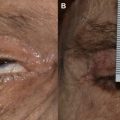
Important overall tenets of facial trauma are to minimize scarring and prevent further injury to adjacent structures. The bony skeleton of the face protects multiple organs that are important to the functions of daily life. It is imperative that these organs are thoroughly evaluated at the initial presentation and the findings accurately documented. Scarring may be unavoidable, depending on the damage to soft tissue from the primary injury and/or location of the fractures and the access required for their repair. Lacerations should be copiously irrigated, minimally debrided, and closed primarily in a layered fashion. Local skin flaps may be used to cover defects, if necessary. Hypertrophic or cosmetically unfavorable scars can be treated with dermabrasion, serial excision, or scar revision.
Brain injuries occur in up to 89% of patients with complex facial trauma. All patients should be evaluated for potential involvement of the brain or cervical spine ( Table 3 ), and an urgent neurosurgical consultation should be obtained for any positive findings. Traumatic brain injuries can be classified as closed, penetrating, and explosive blast injuries, with the severity based on the Glasgow Coma Scale. Cerebrospinal fluid (CSF) leaks carry a 10% to 30% risk of developing meningitis, and can present acutely at the time of initial injury or in a delayed fashion. Symptoms include persistent clear rhinorrhea or otorrhea, description of a salty taste in the mouth by the patient, headaches, or recurrent meningitis, and can be confirmed with a positive β2-transferrin test of collected fluid. Most CSF leaks resulting from accidental and surgical trauma heal with conservative measures over the course of 7 to 10 days, although waiting for the leak to close spontaneously can increase the risk of meningitis, and close assessment to assure that complete resolution has occurred is necessary. Surgical management includes exposure of the leak with primary repair or patch placement. Meningitis is treated aggressively with parenteral broad-spectrum antibiotics. To prevent irreversible neurologic injury, spinal-cord injury should be suspected in all trauma patients until it is ruled out. Repair of facial fractures may initially be delayed while the patient is hemodynamically stabilized. If repair is performed before clearance of the cervical spine, it is imperative that the cervical spine remains in a neutral position. Closed reduction or external fixation techniques may be necessary to avoid injury to the spinal cord if access is inadequate.
| Traumatic brain injury (TBI) | Closed head TBI | Typically a result of blunt impact. May result in a focal lesion in the brain (hematoma) or diffuse axonal injury (from shearing of axons against the skull base) | Neurosurgical consultation necessary |
| Penetrating TBI | Occurs with foreign body violation of the skull and dura, entering the brain parenchyma. The size, speed, and track of the projectile determine the extent of neurologic damage | Neurosurgical consultation necessary | |
| Explosive blast TBI | More common in military combat and causes diffuse injury secondary to a pressure wave. Often results in rapidly developing cerebral edema, subarachnoid hemorrhage, and burst-pattern skull fractures | Neurosurgical consultation necessary | |
| Cerebrospinal fluid (CSF) leak | Symptoms: persistent clear rhinorrhea or otorrhea, description of salty taste in mouth, headaches or recurrent meningitis | Neurosurgical or otolaryngology consultation may be necessary. Conservative management: bed rest, head elevation, avoidance of nose blowing or straining, and stool softeners. Antibiotic prophylaxis and lumbar drain placement is controversial and often surgeon dependent. Current studies have found no benefit from prophylactic antibiotics, though remains controversial Surgical management: transcranial, subcranial, or endoscopic approach with the placement of a mucosal, fascial, or bone graft. The endoscopic approach has become more prevalent and is shown to be safe and effective, with a 90% initial success rate that improves further with subsequent attempts, with lower morbidities than open procedures | |
| Meningitis | Symptoms: headache, nausea, photophobia, altered level of consciousness, fever, nuchal rigidity and pain with flexion of the neck | Empiric first-line treatment in patients with postneurosurgical meningitis is intravenous vancomycin plus cefepime or ceftazidime | |
Approximately 22% to 30% of orbital fractures have associated ocular injuries. It is imperative that all patients are evaluated for vision-threatening injuries and managed emergently to minimize loss of vision ( Table 4 ). The most common vision-threatening injuries include traumatic optic neuropathy, retrobulbar hemorrhage, and penetrating globe injury. Visual acuity, visual fields, color vision, extraocular movement, the pupil, and the fundus should be examined in all patients with periorbital injuries. Diplopia, caused by inflammation and/or edema, is common after both orbital injury and surgery. It may also be evidence of direct injury to the globe, entrapment of orbital soft tissue or extraocular muscles, and vascular or neural damage. Diplopia is usually temporary and should be closely monitored. If persistent after surgical repair, a computed tomography (CT) scan should be obtained to evaluate the implant and fracture repair for misplacement and/or incarceration of soft tissue. Unless entrapment or adherence has been identified, surgical exploration is rarely beneficial, and strabismus surgery may be required. The presence of a retinal injury may preclude immediate repair of periorbital bone injuries, and surgery should be delayed until approved by the consultant ophthalmologist.
| Retrobulbar hematoma (RBH) | Bleeding into the orbit causing increased intraocular pressure compromising the blood supply to the optic nerve and retina. Leads to progressive vision loss and eventual blindness Venous: Slower progression. May not be evident until patient is in recovery or possibly after discharge Arterial: Can progress within seconds, requiring frequent monitoring or palpation of the globe during surgery, especially if significant bleeding is encountered | Symptoms: proptosis, periorbital ecchymosis, increased intraocular pressure, tense globe, loss of direct pupillary light reflex, diplopia, ophthalmoplegia, and decreasing visual acuity/blindness | First-line treatment: Immediate lateral canthotomy and inferior cantholysis Adjunctive treatment: Head of bed elevation or reverse Trendelenburg (cervical-spine precautions), removal of intranasal packing, and immediate ophthalmology consultation with measurement of intraocular pressure. Administration of mannitol 20% (1–2 g/kg IV over 30–60 min), systemic corticosteroids (dexamethasone 8–10 mg IV every 8 h for 3–4 doses), acetazolamide (500 mg IV bolus) or topical antiglaucoma eye drops (Timolol ophthalmic drops 0.5%, 1–2 drops topically twice daily) Second-line treatment: Orbital decompression and anterior/posterior ethmoid artery ligation |
| Traumatic optic neuropathy (TON) | Clinical diagnosis referring to any insult to the optic nerve secondary to trauma Direct TON: penetrating injuries severing the optic nerve. Permanent blindness results Indirect TON: hematoma or secondary edema of the optic nerve within optic canal causing direct mechanical trauma or vascular ischemia, leading to further retinal ganglion cell injury and visual loss | Symptoms: relative afferent pupillary defect in the affected eye and varying loss of visual acuity from partial visual loss to blindness | Ophthalmology consultation is necessary Treatment: No standard of care. Options include observation, corticosteroids, and optic nerve decompression. Spontaneous visual recovery ranges from 0% to 60%. At present neither intervention has been found to be more effective than observation alone. Patients presenting with no perception of light to the injured eye have a poor prognosis for recovery with any course of action |
| Open globe injury/globe rupture | Full-thickness defect of the cornea or sclera | Findings: possible prolapsing uveal tissue, retina, or vitreous gel. Decreased intraocular pressure | Do not manipulate the eye to prevent extrusion of contents. A protective eye shield is placed and emergent ophthalmology consultation is made. Early surgical repair, if possible. Enucleation or evisceration of the globe within 2 weeks with nonsalvageable injuries to avoid sympathetic ophthalmia |
| Sympathetic ophthalmia | Rare, bilateral, granulomatous uveitis presenting after ocular trauma or surgical interventions, causing blindness in the noninjured (sympathetic) eye. Etiology thought to involve inflammatory and autoimmune response after ocular antigens exposed to the immune system. Presents weeks to years after injury | Symptoms: insidious onset of blurry vision, pain, epiphora, and photophobia | Ophthalmology consultation is necessary. Aggressive treatment with systemic corticosteroids or immunosuppressive therapy |
Related posts:

Stay updated, free articles. Join our Telegram channel

Full access? Get Clinical Tree


