(1)
Klinik für Plastische Gesichtschirurgie, Marienhospital Stuttgart, Stuttgart, Germany
Electronic supplementary material
The online version of this chapter (doi:10.1007/978-3-662-49706-7_14) contains supplementary material, which is available to authorized users.
Keywords
Revision rhinoplastyMultiple revised noseSecondary rhinoplastyAdvanced rhinoplastyComplications in rhinoplastySkin necrosisNose reconstructionComplex deformityThe nose is the central aesthetic element of the face, and a failed rhinoplasty may lead to significant psychosocial stress and dysfunction. The impact of a failed rhinoplasty should prompt the surgeon to explore the cause of treatment failure and to consider attempting to salvage the outcome with secondary surgery. However, the inexperienced surgeon often exacerbates the initial treatment failure when attempting to correct his or her own work, creating an ever greater challenge for the next surgeon. In such cases, the patient is often apprehensive, skeptical, and mistrusting of further surgery, even at the hands of an accomplished revision rhinoplasty specialist. However, the embarrassment and emotional angst of a failed rhinoplasty eventually prompt further surgery despite the associated fear and anxiety. Yet, in addition to the psychological issues of the revision rhinoplasty patient, the revision surgeon must also contend with the technical challenges of a previously operated nose. Disruption of surgical navigation planes, excessive bleeding, and dense scarring typically make the dissection far more challenging. Moreover, missing, distorted, or damaged skeletal structures typically necessitate complex skeletal grafting, and graft survival is less reliable due to compromised circulation. In the worst-case scenario, vascular insufficiency can lead to graft or skin necrosis. Even when the technical aspects of surgery are successful, postoperative swelling is often increased, and the healing process is usually prolonged. For all of these reasons, a candid and explicit preoperative discussion in which the limitations, risks, and complications of revision rhinoplasty are reviewed in detail must be undertaken. Additional time must also be allotted for a complete discussion of the relevant issues.
14.1 Case 1: Long-Term Result of Complex Tip Reconstruction (12-Year Follow-Up)
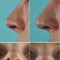
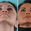
A 50-year-old patient presented following two functional and cosmetic septorhinoplasties. Examination revealed retraction, fixation, and scarring of the supratip skin from previous surgical perforation. Asymmetrical nasal bones with a C-shaped dorsal deformity, overly prominent soft-tissue triangles, and left-sided alar retraction were also observed. Although the open approach carried an increased risk of skin necrosis in the area of iatrogenic skin perforation, the advantage of a precise diagnostic assessment made the open approach justifiable. Upon opening the nose, the left lateral crus and the nasal dome were both missing and had been replaced with a malpositioned cartilage onlay graft. Reconstruction was achieved using donor septal cartilage. Both lower lateral cartilages (LLCs) were reconstructed using narrow strips of septal cartilage thinned with a motorized diamond fraise to create individual domal/lateral crural replacement grafts. Spanning sutures were then used to contour the lobule, and a shield graft fashioned from tragal cartilage was added for additional tip contour. A suspension suture with a posterior sling was then used for tip rotation and stabilization (Figs. 14.1 and 14.2).
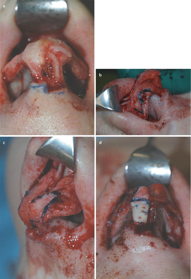
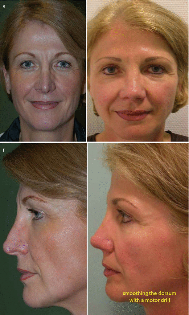
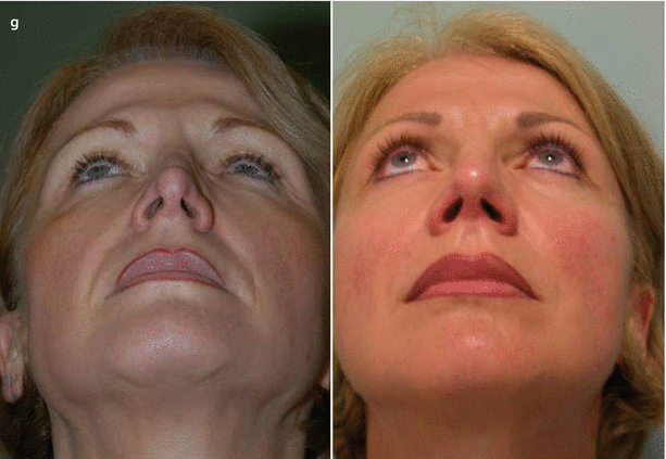
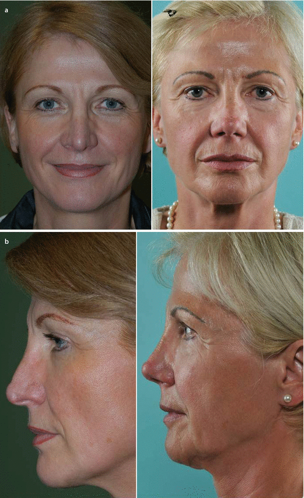
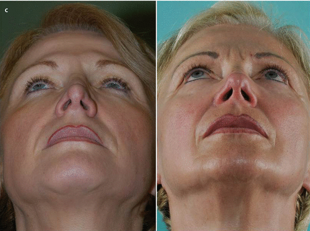
Fig. 14.1
(a, b) Left lateral crus and both domes have been removed and replaced by a combined shield-onlay graft, which has been dislocated. (c, d) Reconstruction of the missing parts by septal cartilage grafts, shield graft from tragus for tip contouring. (c–e) Pre-/postoperative images after 1 year
Fig. 14.2
(a–c) Twelve-year follow-up
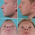
14.2 Case 2: Complex Reconstruction with Extracorporeal Septal Reconstruction, Cartilage Grafts from Ear and Rib and Dorsal Reconstruction with DC-F Graft
A 41-year-old female presented after three previous septorhinoplasties complaining of nasal obstruction and a misshapen nose. Examination revealed an overly short and saddled nose, a pinched tip with retracted soft-tissue facets, and asymmetrical nostrils from a malpositioned columella. Endonasal examination revealed nasal polyposis and an 18-mm septal perforation high in the nasal vault. Open access was achieved via an inverted-V transcolumellar incision located at the narrowest segment of the columella, above the previous malpositioned columellar incision. Dissection was challenging because of extensive surgical scarring, and large portions of quadrangular cartilage were found to be missing, including the caudal septum and the anterior septal angle. Large portions of the LLCs were also previously resected. Reconstruction of the L-strut was accomplished using a double-layered conchal cartilage graft reinforced from behind with small residual strips of septal cartilage sewn to a PDS foil scaffold. The L-strut replacement graft was secured to the nasal spine using a transverse drill hole for suture fixation. The septal perforation was closed using a four-flap technique in which the superior flap was an advancement flap from the upper lateral cartilage mucosa and not a bridging flap, as recommended in some textbooks. The nasal polyps were also removed. Thin and malleable rib cartilage grafts fashioned from the tenth rib were then used to replace the missing LLCs, and a nonintegrated shield graft was also placed. A DC-F graft constructed from autologous temporalis fascia and filled with a combination of diced conchal and rib cartilage was then used to augment the overresected nasal dorsum (Fig. 14.3).
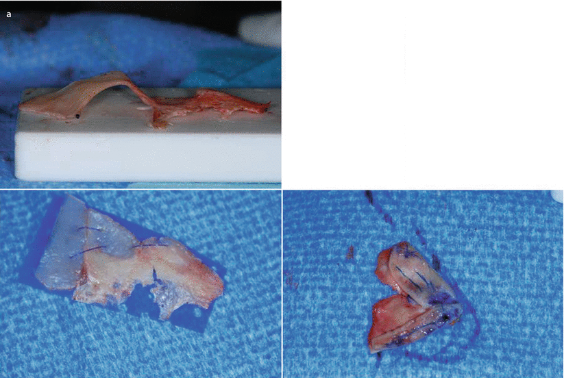
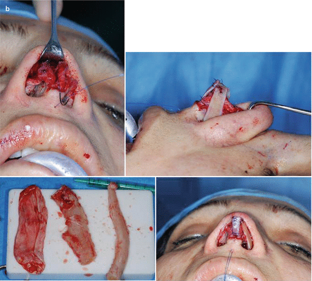
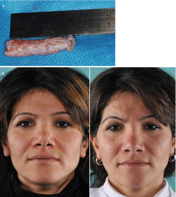
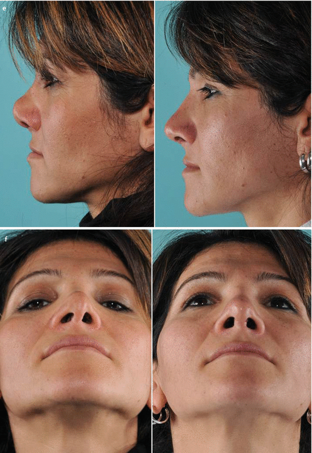
Fig. 14.3
Residual straightened parts of the septum are sutured to a PDS foil. (a) In front of this reconstruction, a double-layered concha graft was placed to reconstruct the anterior septal angle to support the tip and to lengthen the nose. (b) The L-strut replacement graft is brought into position with a translabial guiding suture; rib graft and deep temporalis fascia are harvested; LLCs are reconstructed with strips from rib grafts. (c) DC-F for dorsal reconstruction. (d–f) pre-op/2 yr post-op
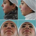
14.3 Case 3: Complex Nose Reconstruction in a Patient with Partial Necrosis of the Columella
A 43-year-old female presented after four previous open rhinoplasties complicated by ischemic necrosis of the columella. Although the primary surgeon recommended immediate columellar reconstruction, we persuaded the patient to wait until the necrotic segment healed by secondary intention. Examination of the nose also revealed a rightward C-shaped deformity of the dorsum, collapse of the right lateral crus, and conspicuous nostril asymmetry. Following healing of the columella, a two-stage revision surgery was recommended in which the columellar scar would be treated secondarily. Open rhinoplasty was performed via a transcolumellar incision located at the upper edge of the depressed columellar scar. Only minor fragments of LLCs remained, and all of the surplus septal cartilage was missing, making septal graft harvest impossible. Although rib cartilage was ultimately used for reconstruction, breast implants, a pectus excavatum, and diffusely calcified rib cartilage made an inframammary crease incision ill-advised. Thin strips of rib cartilage were then fabricated and placed over the LLC remnants. However, owing to excessive cartilage rigidity, transdomal sutures failed to provide a satisfactory tip contour. Consequently, the domes were vertically divided and sutured back together to achieve the desired shape. Dorsal irregularities were then excised, and two layers of allogenic fascia lata were also used for camouflage, for profile augmentation, and for improved dorsal aesthetic lines. Three months later, the patient returned for treatment of the depressed columellar scar. The depressed segment was carefully de-epithelialized and covered with a full-thickness postauricular skin graft (Fig. 14.4).
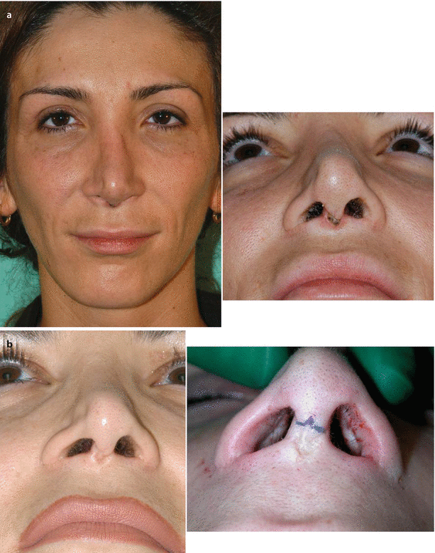
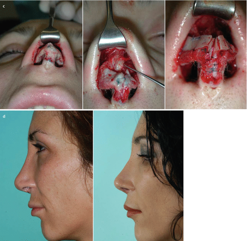
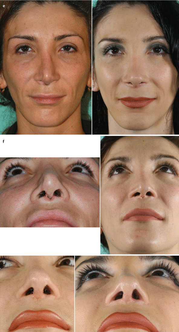


Fig. 14.4
(a) Severe nasal deformity after four previous rhinoplasty, last with open approach, wound healing problems, and partial necrosis of the columellar flap. (b) Deep scar after secondary healing. (c) Reconstruction of the LLCs with rib cartilage (dome division technique). (d–f) Pre-/postoperative images after the first stage of reconstruction (nose). (g) Second stage of reconstruction: correcting the deep retracted columellar scar (f) by full-thickness postauricular skin graft
Related posts:

Stay updated, free articles. Join our Telegram channel
Full access? Get Clinical Tree
