Fig. 3.1
Erythema of the face with accentuation of the ear
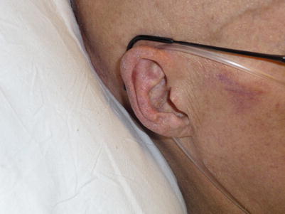
Fig. 3.2
Erythema of the ear
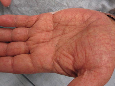
Fig. 3.3
Palmar erythema
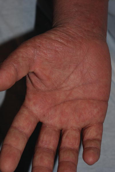
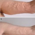
Fig. 3.4
Palmar erythema (Note microvesicles that herald edema.)
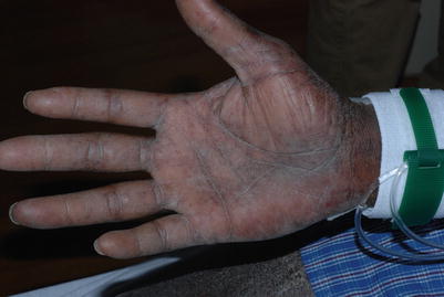
Fig. 3.5
Erythema of the palm
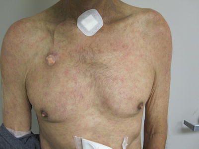
Fig. 3.6
Patchy erythema of the trunk and arms
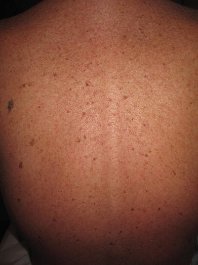
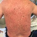
Fig. 3.7
Confluent erythema of the back
Follicular Accentuation
Prominence of hair follicles (whether overlying erythema is present or not) is typical of aGVHD [5]. Such prominence is often misdiagnosed as folliculitis, but it typically heralds the involvement of aGVHD within the follicular epithelium (Figs. 3.8 and 3.9).

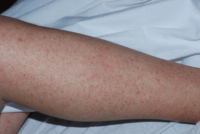
Fig. 3.8
Erythema of the arm with follicular accentuation
Fig. 3.9
Erythema with follicular accentuation of the leg
Morbilliform Lesions
Morbilliform aGVHD is among the most common presentations [6]. Eruptions are erythematous macules and papules that may coalesce into larger papules and plaques, which are symmetric and are often pruritic. This presentation may clinically mimic viral exanthema or drug eruptions (Figs. 3.10, 3.11, 3.12, and 3.13).
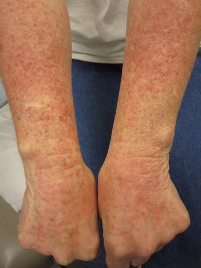
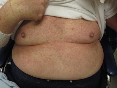
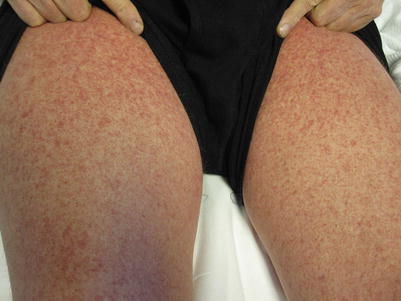
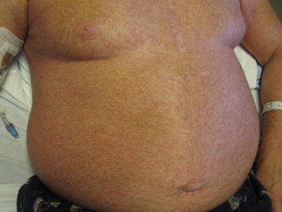
Fig. 3.10
Morbilliform lesions on the dorsal forearms/hands
Fig. 3.11
Morbilliform lesions on the chest and abdomen
Fig. 3.12
Morbilliform lesions on the thighs
Fig. 3.13
Morbilliform lesions diffusely located on the trunk
Erythroderma
Erythrodermic aGVHD presents with confluent erythematous patches that mimic severe viral exanthema, drug reactions, psoriasis, eczematous dermatitis, cutaneous T-cell lymphoma, or staphylococcal scalded skin syndrome (SSSS) (Fig. 3.14). When mucous membrane lesions are present, there are no clinical or histologic differences between skin stage 4 aGVHD and toxic epidermal necrolysis (TEN) [7–9]. If extracutaneous features of aGVHD are present, diagnosis may be possible via tissue confirmation at involved sites. If extracutaneous features of aGVHD are absent, review of clinical symptoms and their development in relation to initiation of medications is mandatory. Viral serologies and/or quantification of viremia via polymerase chain reaction (PCR) testing of the blood may aid in the diagnosis of an erythrodermic viral exanthem, such as those caused by HHV-6. Concurrent empiric treatment of aGVHD, viral infection, and TEN may be necessary if a clear diagnosis cannot be rendered.
 Grading and Treatment of Acute Graft-Versus-Host Disease
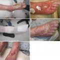 Wound Care in the Management of Chronic Graft-Versus-Host Disease
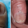 Clinical Presentation of Nonsclerotic Epidermal Chronic Graft-Versus-Host Disease and Hair and Nail Changes
 Dermal and Subcutaneous Chronic Graft-Versus-Host Disease
 Diagnosis, Staging, and Treatment of Chronic Graft-Versus-Host Disease
 Clinical Presentation of Nonsclerotic Epidermal Chronic Graft-Versus-Host Disease and Hair and Nail Changes


Related posts:
Grading and Treatment of Acute Graft-Versus-Host Disease
Wound Care in the Management of Chronic Graft-Versus-Host Disease
Clinical Presentation of Nonsclerotic Epidermal Chronic Graft-Versus-Host Disease and Hair and Nail Changes
Dermal and Subcutaneous Chronic Graft-Versus-Host Disease
Diagnosis, Staging, and Treatment of Chronic Graft-Versus-Host Disease
Clinical Presentation of Nonsclerotic Epidermal Chronic Graft-Versus-Host Disease and Hair and Nail Changes
Stay updated, free articles. Join our Telegram channel
Full access? Get Clinical Tree
