Cleft Lip and Palate: Embryology, Principles, and Treatment


Cleft Lip and Palate: Embryology, Principles, and Treatment


Richard A. Hopper


Cleft lip and palate are the most common congenital craniofacial anomalies. Successful treatment requires technical skill, knowledge of the abnormal anatomy, and appreciation of three-dimensional facial aesthetics. Cleft care requires a collaborative multidisciplinary team. Through self-scrutiny, honest evaluation of the results, and a great deal of imagination, plastic surgeons continue to advance cleft care.


EMBRYOLOGY


Developmental Biology

An understanding of head and neck embryology is helpful in the appreciation of the wide spectrum of the cleft lip and palate phenotype. The cranial portion of the human embryo develops early, with the three germ layers (ectoderm, mesoderm, and endoderm) forming in the beginning to middle of the third week of gestation. The ectoderm layer gives rise to the cutaneous and neural systems, with differentiation starting at 20 days. The interaction between ectoderm-derived components at the crest of the neural fold gives rise to a unique cell population of neural crest cells (NCCs). NCCs have the unique ability to remain pluripotent despite their single germ layer origin. NCCs migrate along cleavage planes between germ layers and within the mesoderm to differentiate at their final destination into connective, muscle, nervous, or endocrine tissue, as well as pigment cells.

NCCs that migrate ventro-caudal from the crest come into contact with the pharyngeal endoderm and mesoderm core that surrounds the six aortic arches. This results in a series of mesenchymal swellings termed branchial arches in the fourth week. The six paired branchial arches decrease in size from cranial to caudal. Although the first and largest arch, the mandibular arch, is primarily responsible for development of the anatomy that includes the lip and palate, the fourth arch is responsible for the pharyngeal constrictor, the levator veli palatini, and the palatoglossus muscles, which play a role in the problems and treatments associated with cleft palate. Each branchial arch gives rise to a nerve along with the associated muscles. This muscle-nerve relationship is maintained regardless of the functional interaction of the differentiated structures. Although the tensor veli palatini and levator veli palatini work in close coordination in the mature normal palate and are pathologically tethered through the aponeurosis of the tensor tendon in patients with cleft palate, they retain their distinct innervation based on their embryologic origin. The levator veli palatini, as a fourth arch derived muscle, is innervated by the fourth arch derived superior laryngeal branch of the vagus (cranial nerve X). The fourth arch derived palatoglossus and pharyngeal constrictors have similar innervation. The tensor veli palatini alone, as a derivative of the first arch, is innervated by the trigeminal nerve (cranial nerve V).

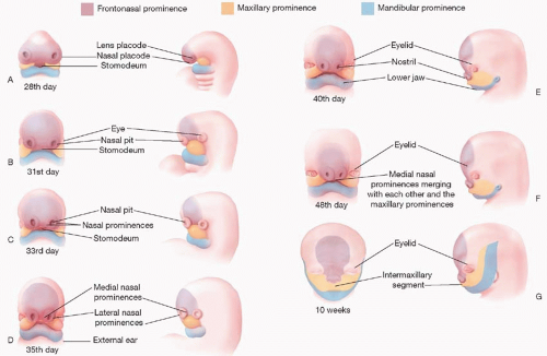
The first branchial arch and the mesenchyme ventral to the developing forebrain are responsible for the three named prominences that give rise to the face, mouth, neck, larynx, pharynx, and nasal cavities (Figure 19.1). The first branchial arch contributes the paired maxillary and mandibular prominences, which fuse to form the lateral and caudal components of the primitive stomodeum or mouth. A central process formed by the proliferation of mesenchyme ventral to the forebrain creates the frontonasal prominence (FNP), which forms the cranial portion of the stomodeum. It is important to note that the FNP and its derivatives are not formed by branchial arches, but rather originate from distinct mesenchyme ventral to the branchial arches. These five facial prominences (two paired and one unpaired) are separated by external grooves, but the mesenchyme of all five is continuous, such that unobstructed migration of mesenchymal cells can occur around the stomodeum. Coordinated fusion and communication between these five prominences are essential for normal lip and palate development.

Development of the human face occurs between the 4th and 10th weeks (Figure 19.1B-H). The nasal placodes develop as bilateral thickenings on the surface ectoderm of the infero-lateral aspect of the FNP by the end of the fourth week (Figure 19.1C). As the placodes elevate, medial and lateral nasal prominences develop around the depressed central nasal pit. Medial migration of the maxillary prominence from the first arch effects medial migration of the nasal prominences, such that when they fuse together, the stomodeum is no longer in continuity with the nasal pit, creating the nasal-oral separation (Figure 19.1G).

The medial nasal prominences form the philtrum and Cupid’s bow region of the upper lip, the nasal tip and septum, and the premaxilla back to the incisive foramen. The lateral nasal prominences form the nasal alae. The maxillary prominences form the lateral lip elements that normally fuse with the philtrum derived from the medial nasal prominence. A failure of fusion of a lateral lip element (maxillary prominence) with the philtrum (medial nasal prominence from the FNP) results in a unilateral cleft lip. If both maxillary prominences fail to fuse, a bilateral cleft lip will result. With a failure of fusion to the maxillary prominence, the growth of the medial placode elements (prolabium, premaxilla, and septum) is unbalanced, resulting in the central protrusion seen in a cleft patient.

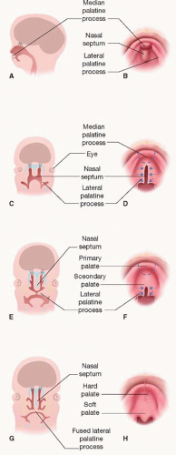
The formation of the palate is also a result of interaction between the FNP and maxillary prominences. The two medial nasal prominences of the FNP merge to form the median palatine process, which develops the primary palate, whereas the
lateral palatine processes derived from the maxillary prominences form the secondary palate (Figure 19.2). During the eighth week, the lateral palatine processes change from their initial vertical orientation to horizontal, within a period of hours. The developing mandible protrudes in synchrony to allow the tongue to descend and leave room for palate fusion. Fusion occurs in both the axial and sagittal planes, with the median palatine process and two lateral palatine processes fusing to form the palate, and the nasal septum descending from the FNP to join the fusion and separate the two nasal cavities (Figure 19.2C-F). Fusion involves focal degeneration of the leading epithelial edges in a process felt to represent “programmed cell death.” Once fused, the mesenchyme of the primary palate and anterior secondary palate ossify into the hard palate, whereas the posterior secondary palate forms muscle to create the dynamic soft palate.


FIGURE 19.1. Illustrations of the progressive stages of the development of the human face. From gestational age of 28 days (A) through 10 weeks (G) there is staged and progressive fusion of the frontonasal (purple), maxillary (orange) and mandibular (blue) prominences.

When there is normal fusion between the FNP and maxillary prominences creating a normal lip and alveolus, but there is lack of fusion between the lateral palatine processes of the opposing maxillary prominences, an isolated cleft of the secondary palate occurs. If, however, the maxillary prominences fuse appropriately, creating a normal secondary palate, but the FNP and maxillary prominences do not fuse, then a cleft lip and cleft of the primary palate will occur. The variety of fusion patterns between these two pathologic scenarios results in the plethora of cleft lip and palate combinations described later in the chapter.


Epidemiology and Etiopathogenesis

Among the cleft lip and palate population, the most common diagnosis is cleft lip and palate (46%), followed by isolated cleft palate (33%) and isolated cleft lip (21%). The majority of bilateral cleft lips (86%) and unilateral cleft lips (68%) are associated with a cleft palate. Unilateral clefts are nine times as common as bilateral clefts and occur twice as frequently on the left side than on the right. Males are predominant in the cleft lip and palate population, whereas isolated cleft palate occurs more commonly in females. In the Caucasian population, cleft lip with or without cleft palate occurs in approximately 1 in 1,000 live births. These entities are twice as common in the Asian population, and approximately half as common in African Americans. This racial heterogeneity is not observed for isolated cleft palate, which has an overall incidence of 0.5 per 1,000 live births.

Both environmental teratogens and genetic factors are implicated in the genesis of cleft lip and palate. Intrauterine exposure to the anticonvulsant phenytoin is associated with a 10-fold increase in the incidence of cleft lip. Maternal smoking during pregnancy doubles the incidence of cleft lip. Other teratogens, such as alcohol, anticonvulsants, and retinoic acid, are associated with malformation patterns that include cleft lip and palate, but have not been directly related to isolated clefts.

Genetic abnormalities can result in syndromes that include clefts of the primary or secondary palates among the developmental fields affected. More than 40% of isolated cleft palates are part of malformation syndromes, compared with less than 15% of cleft lip and palate cases. The most common syndrome associated with cleft lip and palate is van der Woude syndrome with or without lower lip pits or blind sinuses. Microdeletions of chromosome 22q resulting in velo-cardiofacial, DiGeorge, or conotruncal anomaly syndromes are the most common diagnoses associated with isolated cleft
palate. Although there is a recognized genetic component to nonsyndromic cleft lip and/or palate, it appears to be multifactorial. Among other recent studies, a meta-analysis of 13 genome scans by Marazita et al.1 revealed multiple cleft lip/palate genes on 16 chromosomal regions.


FIGURE 19.2. A. Sketch of a sagittal section of the embryonic head at the end of the sixth week showing the median palatine process, or primary palate. B, D, F, and H. Drawings of the roof of the mouth from the 6th to 12th weeks illustrating development of the palate. The broken lines in (D) and (F) indicate sites of fusion of the palatine processes. The arrows indicate medial and posterior growth of the lateral palatine processes. C, E, and G. Drawings of the frontal sections of the head illustrating fusion of the lateral palatine processes with each other and the nasal septum, and separation of the nasal and oral cavities.

Parents of a child with a nonsyndromic cleft, or a family history of clefting, always ask about the risk of clefts in subsequent pregnancies. The risk depends on whether the proband has a cleft lip alone (CL), cleft lip with cleft palate (CLP), or a cleft palate alone (CP). If the family has one affected child or parent with CLP, the risk of the child of the next pregnancy having CLP is 4%. If two previous children have CLP, the risk increases to 9%, and if one parent and one child were previously affected, the risk to children of subsequent pregnancies is 17%. For families with a child having CP, the risk of CP to children of subsequent pregnancies is 2%, 6% if one parent has CP, and 15% if one parent and one previous child have CP.


PRINCIPLES


Surgical Evaluation and Classification

The newborn infant with a cleft is ideally evaluated by the cleft team in the first weeks of life. The increasing number of clefts detected by prenatal imaging allows early preparation of the family and introduction to the treatment plan. Patients with cleft lip and/or palate are not a homogenous group. As mentioned above, they can be divided into CL, CP, and CLP; however, the surgical treatment plan requires a more complex classification scheme. The cleft lip deformity is typically divided into unilateral or bilateral, and then subdivided into complete, incomplete, or microform based on the pattern of embryonic fusion described earlier. The width of the cleft deformity and the degree of alveolar arch collapse also play a part in surgical planning, as these directly relate to the degree of associated nasal deformity and the tension and difficulty of the repair. The associated nasal deformity is similarly categorized as mild, moderate, or severe. Mild nasal deformity is characterized by a lateral displacement of the alar base with normal alar contour, minimal columella shortening, and normal dome projection. Moderate nasal deformity has lateral and posterior displacement of the alar base, columella deficiency, and a depressed dome with mild separation of the interdomal space. Severe nasal deformity has an underprojecting alar dome with complete collapse of the lower lateral cartilage and a severe deficiency of columella height with a dramatic interdomal separation. Severe nasal deformities often have a reversed curvature to the alar rim. The nasal deformity is secondary to a three-dimensional distortion of the lower lateral cartilage, described by some as the “tilted tripod.” It is not caused by hypoplasia or deficiency of the cartilage itself.

If a cleft palate is present, it is surgically classified as unilateral, bilateral, or submucous. A submucous cleft results from epithelial fusion of the soft palate, but lack of the programmed mesenchymal fusion described in the embryology section. The width of the cleft is noted as it affects the difficulty of closure.

Although most surgeons use the descriptive classification of cleft deformities during the initial assessment of a patient, other classification systems are often used for outcome research and record keeping. Kernahan and Stark’s “stripped Y” diagrammatic classification scheme and its modifications continue to be used in many cleft centers. It recognizes the embryologic division of the primary (lip and alveolus) and secondary palates at the incisive foramen. Kriens presented a palindromic acronym organization of cleft deformities. The acronym LAHSHAL denotes the bilateral anatomy of lip (L), alveolus (A), hard (H), and soft (S) palates, by convention from right to left. Lowercase letters represent incomplete clefts of the structure; a period denotes no cleft. A bilateral cleft lip with a complete unilateral cleft of the secondary palate, with incomplete clefting of the lip and alveolus on one side would be represented as LAHSal. This system is currently used for the outcomes registry of the American Cleft Palate and Craniofacial Association (ACPA).


Microform Cleft Lip.

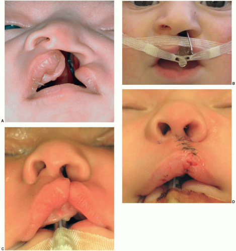
The microform cleft (Figure 19.3A) is characterized by a furrow or scar transgressing the vertical length of the lip, a vermilion notch, imperfections in the white roll, and varying degrees of vertical lip shortness. Nasal deformity may be present and is sometimes more extensive than the associated deformity of the lip. Surgery is generally indicated but is approached cautiously to avoid a surgical deformity worse than the congenital defect. If there is isolated disruption of the orbicularis oris sphincter, it can be repaired through an intraoral approach.


Unilateral Incomplete Cleft Lip.

Unilateral incomplete clefts (Figure 19.3B) are characterized by varying degrees of vertical separation of the lip, but they all have in common an intact nasal sill. They typically require the same surgical technique as a complete cleft lip in order to repair the underlying muscle malposition, with the associated distortion of the septum, alar base, and lip. If the nasal sill skin is normal, and the nasal lining intact, one of the challenges of the incomplete cleft is to elevate the nasal lining from the underlying alveolar cleft to allow repositioning of the alar base while preventing a nasolabial fistula. As with complete clefts, the best time to address the associated nasal and septal deformity is at the time of the primary lip repair.


Unilateral Complete Cleft Lip.

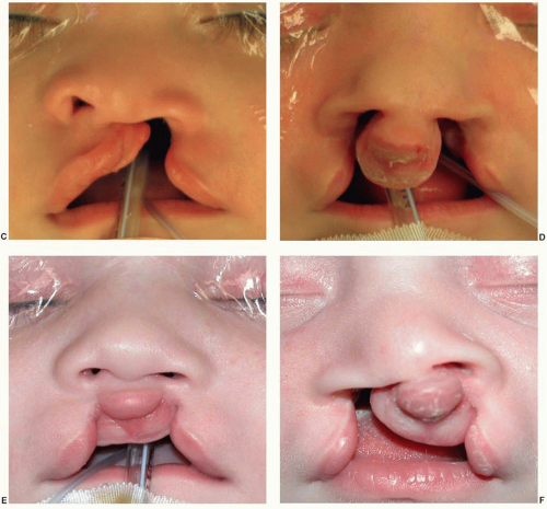
Unilateral complete clefts (Figure 19.3C) are characterized by disruption of the lip, nostril sill, and alveolus (complete primary palate). Since there is no skin bridge connecting the alar base to the footplates of the lower lateral cartilages of the nose, unopposed pull of the orbicularis oris muscle results in a more severe nasal deformity than seen in an incomplete cleft lip. The alar base is displaced inferior and posterior, the ipsilateral lower lateral cartilage of the nose is stretched and the natural contour deformed, and the floor of the nasal septum is displaced into the non-cleft nostril, collapsing the nasal tip support. The critical factors for evaluating unilateral complete clefts are the position of the lesser and greater alveolar segments, the vertical height of the lateral lip element, and the degree of associated nasal deformity. The alveolar (maxillary) segments assume one of four positions: (a) narrow-no collapse; (b) narrow-collapse; (c) wide-no collapse; (d) wide-collapse. “Wide” is determined by an alveolus position lateral to the desired alar base position (i.e., with lip closure the alar base is medial to the alveolus and thereby sitting in the cleft). “Collapse” refers to a palatal displacement of the lateral maxillary segment as predicated by the arch configuration of the medial, non-cleft dental ridge.


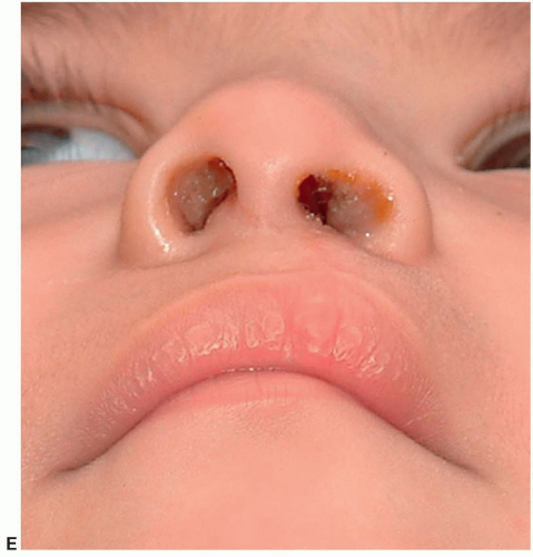
FIGURE 19.3. The clinical spectrum of cleft lip deformities. A. Microform cleft lip. B. Unilateral incomplete cleft lip. C. Unilateral complete cleft lip. D. Bilateral complete cleft lip. E. Bilateral incomplete cleft lip. F. Hybrid incomplete and complete bilateral cleft lip.

Clefts characterized as “narrow-no collapse” with minimal nasal deformity may be treated with presurgical taping to prevent widening of the cleft with growth and feeding, prior to a primary cleft lip repair with primary tip rhinoplasty. Clefts characterized as “narrow-collapse” or “wide-collapse” may benefit from presurgical molding to create the desired arch form, alveolar contact, and nasal anatomy at the time of surgery. Clefts characterized as “wide-collapse” or “wide-no collapse” must be assessed closely by the dental members of the cleft team. If they feel that these cases are deficient in arch mesenchyme, presurgical orthopedics is used to align the arch segments by correcting the collapse, but not to close the alveolar cleft since this will result in a constricted or perhaps locked in arch. External taping can be used to correct the alar base position over the maintained arch form. The use of presurgical orthopedics or aggressive presurgical taping has eliminated the need for preliminary lip adhesion surgery. The primary benefit of a balanced arch configuration at the time of primary lip repair is decreased tension on the lip repair. A secondary benefit is the reduction of alar discrepancy.


Complete Bilateral Cleft Lip.

The most obvious aspect of a complete bilateral cleft is the protruding premaxilla (Figure 19.3D). Because of the lack of connection of the premaxilla with the lateral palatal shelves, the premaxilla has not been “reined back” into alignment with the lateral arch segments during fetal development. At the time of birth, the premaxilla protrudes on a vomerine stem. Uncontrolled growth at the premaxillary suture results in over-projection of the premaxilla, with or without rotation and angulation of the segment. Just as the premaxilla is not reined back by the lateral palatal shelves, the lateral palatal shelves are not pulled forward by their attachment to the premaxilla. Without the intervening premaxilla to maintain arch width, the lateral palatal shelves collapse toward the midline. The severity of this disruption of arch morphology varies, and will dictate the tension on the repair, the degree of dissection required, and,
ultimately, the final aesthetic result unless it is corrected prior to lip repair. Presurgical orthopedics is employed to achieve this correction prior to surgery.


FIGURE 19.3. (Continued)

The anterior nasal spine is poorly formed or absent in the bilateral cleft lip deformity, resulting in a retruded area under the base of the septal cartilage and recession of the footplates of the medial crura. The footplates of the lower lateral cartilages are displaced posterior and laterally, which in turn pulls the normal junction (genu) of the medial and lateral crura apart resulting in a broad, flat nasal tip. The recession of the medial crural footplates along with lateralization of the domes and deficient skin produces the typical “absent columella” deformity. The most anterior and inferior extent of the frontonasal process, which normally contributes to the skin between the philtral columns of the lip, forms a wide, short disk, called a prolabium, that appears to hang directly from the nasal tip skin. In conventional techniques, the linear distance from the inferior tip of the prolabium to the nasal tip is inadequate to reconstruct both the central upper lip and columella length. This vertically limited tissue is used to create the central lip element at the cost of inadequate columella length and tip projection. A major benefit of nasoalveolar molding (NAM) is the ability to lengthen both the columella skin and the prolabium prior to surgery, creating enough skin to reconstruct the central lip length without compromising nasal tip projection.


Incomplete Bilateral Cleft Lip.

Occasionally, bilateral clefts are incomplete with a near-normal nose, a normally positioned premaxilla, a skin bridge across one or both nasal floors, and clefts involving only the lip (Figure 19.3E). In such circumstances, a rotation-advancement approach, or a triangular flap approach similar to that used in unilateral repairs, can be used either in a single-stage or a two-stage operation. In two-stage repairs one side is closed first, allowed to heal, and then the other side is repaired a short time later. Symmetry is difficult to achieve with a staged approach, and we prefer a single-stage procedure with a bilateral straight-line technique as described later in the chapter. Patients with a complete cleft on one side and an incomplete cleft on the other
present a surgical challenge (Figure 19.3F). These cases have the asymmetric nasal deformity of a unilateral complete cleft lip and the paucity of lip tissue of a bilateral cleft. If there is a discrepancy in columella height between the two sides, we will consider a rotation-advancement repair on the complete side to increase columellar length and a straight-line closure on the incomplete side.


Cleft Lip and Palate

The primary palate consists of the lip, alveolus, and anterior palate back to the incisive foramen. The secondary palate consists of the hard and soft palates from the incisive foramen back to the uvula. The presence of a cleft palate introduces feeding difficulties, concerns regarding speech development, and the possibility of impaired facial growth. The width of a primary palate cleft and the degree of collapse are typically increased in the presence of a cleft of the secondary palate. The family is counseled about the increased number of surgical operations that will be required if a cleft palate is present: primary cleft palate repair with intravelar veloplasty; possible secondary surgery on the palatopharyngeal muscle sling, such as a sphincteroplasty or pharyngeal flap; and possible orthognathic surgery at skeletal maturity. The abnormal attachment of the muscles of the soft palate in a cleft palate alters the tension on the pharyngeal drainage of the Eustachian canal, increasing the incidence of ear infections. Myringotomy and grommet tube placement is performed in the majority of infants at the time of either the lip repair or the palate repair to prevent the development of hearing abnormalities.


Isolated Cleft Palate

The infant with isolated cleft palate is examined carefully for manifestations of the Pierre Robin sequence (micrognathia and glossoptosis leading to airway obstruction). The cause of the cleft palate in the Pierre Robin sequence is thought to be mechanical obstruction of the lateral palatine processes as they swing from a vertical to horizontal orientation during palate fusion, and not because of failure of the fusion process secondary to “programmed cell death.” The micrognathia and associated glossoptosis causes this obstruction, resulting in the characteristic wide “horseshoe” cleft palate. Since the associated findings are caused by a “domino effect” starting with the micrognathia, and not a shared etiology, the condition is considered a sequence and not a syndrome. If the Pierre Robin sequence is present, the majority of cases can be treated with positioning and anti-reflux medications. In more severe cases, treatment may include nasopharyngeal airway protection, gavage feedings, and apnea monitoring. A small percentage of Pierre Robin patients require surgical intervention such as tongue-lip adhesion, distraction lengthening of the mandible, or tracheostomy. Because of airway concerns, palatoplasty may be delayed for several months in Pierre Robin patients compared with other cleft palate closures.


Submucous Cleft Palate

The submucous cleft palate is traditionally defined by a triad of deformities: a bifid uvula, absence of the posterior nasal spine resulting in a notched posterior hard palate, and muscular diastasis of the velum resulting in a zona pellucidum. Submucous clefts vary considerably, however, and muscular diastasis can occur in the absence of a bifid uvula. The majority of patients with submucous cleft palate are asymptomatic. Approximately 15% of patients will develop velopharyngeal insufficiency (VPI). VPI correlates with short palatal length, limited mobility, and easy fatigability of the palate. Because the majority of patients with submucous cleft palate remain asymptomatic, a non-operative approach is recommended until speech can be adequately evaluated, which is typically after 3 years of age.


Jun 26, 2016 | Posted by in General Surgery | Comments Off on Cleft Lip and Palate: Embryology, Principles, and Treatment

Full access? Get Clinical Tree

Get Clinical Tree app for offline access