CHAPTER 27 Indocyanine Green Lymphography
KEY POINTS
Indocyanine green (ICG) lymphography, which can visualize superficial lymph flow in real time without radiation exposure, is a clinically useful evaluation method for lymphedema.
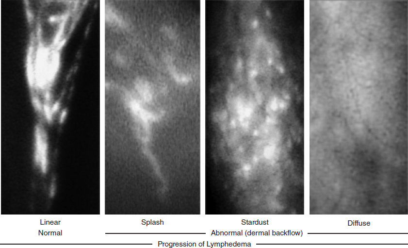
With progression of lymphedema, the ICG lymphographic pattern changes from the normal linear pattern to abnormal dermal backflow (DB) patterns (splash, stardust, and diffuse patterns).
ICG velocity and lymph transportation capacity decrease as lymphedema progresses.
ICG lymphography is also useful for the preoperative assessment of lymphatic surgeries, because different ICG lymphographic findings represent different conditions of the lymphatic vessels; the more severe the DB pattern seen on ICG lymphography, the more sclerotic and smaller the lymphatic vessels are.
In dynamic ICG lymphography, one ICG injection is enough for pathophysiologic severity staging (DB stage), lymph pump function evaluation (ICG velocity), and preoperative assessment.
Several methods have been reported to visualize lymph flow and evaluate lymphedema, including MRI, CT, ultrasonography, and lymphoscintigraphy. 1 – 4 Currently, lymphoscintigraphy is considered the benchmark for the evaluation of lymphedema. Lymphoscintigraphy can visualize deep lymphatic flow but its image is obscure, and it has a risk of radiation exposure. 4 , 5 ICG was first reported in 2007 for the evaluation of lymphedema. 6 , 7 ICG lymphography allows much clearer visualization of superficial lymph flow than lymphoscintigraphy. It is useful not only for lymphedema evaluation but also for the preoperative assessment of lymphatic surgeries. 6 – 16 ICG lymphography is increasingly used in the clinical practice of lymphedema management.
Material for Indocyanine Green Lymphography
As an optical tracer agent, ICG is a medically useful green dye that was approved by the FDA in 1956. ICG is not only a green dye marker but also a fluorescent substance. Near-infrared fluorescent light penetrates tissue more deeply than does visible light. 6 , 7
ICG lymphography, which is used to visualize superficial lymph flow after injection of ICG, is performed with a nearinfrared camera device that is equipped with a charge-coupled device camera as a detector with a 760 nm light-emitting diode and a filter-cutting light below 820 nm. 6 – 12 The fluorescent images are digitized for real-time display by using a standard personal computer. Several nearinfrared camera devices are available. Some manufacturers include Hamamatsu Photonics, Mizuho, Novadaq Technologies, Olympus (microscope), Carl Zeiss (microscope), and Leica (microscope). 13 , 14
Because ICG lymphography is used to visualize lymph flow in real time, ICG lymphography allows static evaluation of lymph circulation as well as dynamic lymph movement. With this property, dynamic ICG lymphography, or dual-phase ICG lymphography, was developed for comprehensive assessment of lymphedema. 17 , 18
Procedures of Dynamic Indocyanine Green Lymphography
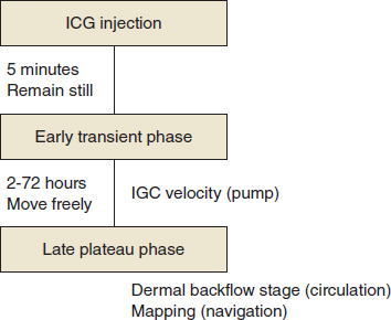
Dynamic ICG lymphography is performed as follows. After a patient remains still for 15 minutes, 0.05 to 0.2 ml of ICG (Diagnogreen 0.25%, Daiichi Pharmaceutical, Tokyo, Japan) is injected subcutaneously (at the second web space for extremity and genital lymphedema and at the glabella and below the nose for facial lymphedema). 8 – 12 Immediately after ICG injection (transient phase), fluorescent images of lymphatic flow are obtained with an infrared camera system. 17 , 18 The observation is continued until lymph pump function is measured. Patients remain still in the supine position during lymph pump function measurement. Then patients are allowed to move freely.
Twelve to 18 hours after ICG injection, ICG movement usually reaches the plateau phase. In this phase, lymph circulation is evaluated based on ICG lymphographic findings, which allow pathophysiologic severity staging and preoperative guidance for lymphatic surgeries. 8 – 12 When patients move their extremity rigorously, ICG can reach a plateau 2 hours after ICG injection. 17 , 18 The plateau phase usually continues until 72 hours after ICG injection. Thus it is possible to evaluate abnormal lymph circulation between 2 and 72 hours after ICG injection. 15 , 16
In dynamic ICG lymphography, lymphatic images are taken twice; at an early transient phase and at a late plateau phase. 17 , 18 We usually inject ICG the day before lymphatic surgery; one ICG injection is enough for lymph pump function measurement, evaluation of abnormal lymph circulation, and preoperative mapping (Fig. 27-1).
Evaluation of the Lymphatics
LYMPH VESSELS
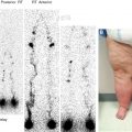
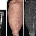
As ICG lymphographic patterns change according to the pathophysiologic changes of lymph flows, different ICG lymphographic findings indicate different conditions of the lymphatic vessels. 9 – 16 As ICG lymphographic patterns change from linear to splash, stardust, and diffuse pattern, lymphatic vessels change to become more sclerotic with less lymph flow. Lymphatic vessels are almost intact in linear regions; on the other hand, they are very sclerotic with a pinhole-like lumen in diffuse regions 9 – 14 (Fig. 27-2). According to a comparative study of ICG lymphographic findings and intraoperative conditions of 215 lymphatic vessels, the mean diameter of the lymphatic vessels was 0.45 mm in linear regions, 0.44 mm in splash and stardust regions, and 0.26 mm in diffuse regions. 15 In planning lymphaticovenular anastomosis, diffuse regions should be avoided, because the detection rate of lymphatic vessels favorable for lymphaticovenular anastomosis is low. 15 , 16
LYMPH TRANSPORTATION CAPACITY
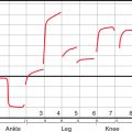
Because ICG lymphography visualizes lymph flow in real time, lymph transportation capacity can be directly evaluated by measuring ICG movement at an early transient phase. There are several ways to evaluate lymph pump function, such as transit time, lymphatic pressure, and ICG velocity. 17 – 20 Among the various lymph pump function evaluations, ICG velocity is the most practical one. ICG velocity can be measured within 5 minutes, whereas others sometimes require more than 1 hour in patients with severe lymphedema. The distance between the injection point and farthest proximal point at which the dye can be observed is measured 5 minutes after ICG injection; ICG velocity is calculated by dividing the distance by time. When ICG reaches the axilla and groin within 5 minutes after dye injection in patients with extremity lymphedema, ICG velocity is calculated by dividing the distance between the injection point and axilla and groin by the time required. ICG velocity decreases with the progression of lymphedema and increases after successful interventions. Because ICG velocity is a quantitative evaluation, it is easy to evaluate lymph pump function before and after therapeutic interventions.
Related posts:
Stay updated, free articles. Join our Telegram channel

Full access? Get Clinical Tree


