Chapter 19 STRUCTURAL FAT GRAFTING FOR THE REGENERATION OF IRRADIATED TISSUE
Evidence of the dynamic and regenerative nature of grafted fat has been demonstrated in several plastic surgery applications as well as in molecular and cell biology studies. 1 Several clinical reports have shown that fat grafting definitively has positive long-term effects both for rejuvenation and regeneration.
Cell biology has shown that the plasticity of adult fat results from the presence of multipotent stromal populations endowed with immunophenotypical and differentiative properties that are similar, but not identical, to the properties of bone marrow–derived mesenchymal stem cells (BM-MSCs). Twenty years ago, experimental studies of autologous fat cell biology revealed the presence of fibroblast-like mesenchymal cells that were able to survive when cultured, to proliferate, and even to differentiate into adipocytes. 2 In addition, recent studies have shown that the stromal-vascular cell fraction of adipose tissue provides a rich reservoir of regenerative precursor cells with proangiogenic capabilities. 3 – 5 In animal models, adipose stromal cells were proven to secrete angiogenic and antiapoptotic factors, 6 and to differentiate into endothelial cells and incorporate into vessels, 7 thus promoting neovascularization in ischemic tissues.
Material and Methods
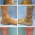
Irradiation-induced lesions are well-known side effects of external oncologic radiotherapy; this is a progressive problem that does not improve over time; on the contrary, it tends to worsen with time. That is why the most severe sequelae, such as skin ulcers and osteoradionecrosis, occur years after radiotherapy. We previously demonstrated that radiation produces an inflammatory reaction causing hyperpermeability of the capillary vessels, increased perivascular edema, and consequent progressive occlusion of the blood vessels. 8 These processes lead to an altered and reduced blood flow, resulting in ischemic damage. 9 Different methods have been proposed for treatment of chronic ischemic radiolesions, with varying degrees of success. Our previous pilot study was conducted to evaluate the regenerative capabilities of adipose-derived adult stem cells contained in lipoaspirates and their potential role in ischemic tissue regeneration. 8 The beneficial effects of fat grafting on these radiation-induced lesions were striking and were generalized to the entire patient population; these effects are now supported by long-term follow-up. Significant improvements were observed in patients with cutaneous ulcers and osteoradionecrotic complications.
The clinical results we achieved in these patients, combined with the extreme accessibility of lipoaspirate obtained by minimally invasive procedures and the abundance of fatty tissue in the body, indicate that structural fat grafting for ischemic tissue regeneration is a particularly effective technique, with great potential for use in routine clinical practice. 10
INDICATIONS
Fat grafting is indicated for patients with progressive lesions after radiotherapy. These patients are usually screened and objectively assessed based on the late effects in normal tissues per subjective, objective, management, and the analytic (LENT-SOMA, or LS) scale, 11 , 12 where grade 1 corresponds to light symptoms and grade 4 corresponds to irreversible functional damage. There are no specific reasons for patient exclusion from therapy because of his or her LS score. Therapy is recommended when symptoms are classified LS grade 2, including fibrosis, atrophy, retraction, ulcers, and telangiectasias, accompanied by itching and pain. There are no specific limitations related to patient age and radiolesion site, but the breast, with and without an expander or a prosthesis, is the most frequent site. Therefore the administered radiation dose, time elapsed since radiation treatment, and the severity of symptoms associated with the radiolesion (LS score) do not represent contraindications to fat grafting. The elapsed time since the end of radiotherapy and the first fat grafting session greatly varied in our series, from 6 months to 44 years. Fat grafting to the irradiated breast should usually be performed at least 6 to 8 months after the resolution of the immediate side effects of radiation.
TECHNICAL GUIDELINES
Preparation
Preoperatively, the targeted area should be precisely identified and the number and locations of entry points and direction of tissue injection tunnels defined; these parameters can be refined during surgery. The goal is to achieve maximal uniformity of distribution and to limit overlaps and gaps in tissue deposition.
Harvest
A local anesthetic is used for harvesting fatty tissue, with the patient under deep sedation. Antibiotics are administered only once, 20 minutes before the procedure. Typical donor sites include the medial aspect of the knee, the abdomen, or the trochanteric areas, because these are frequently a rich reservoir of fatty tissue. The selected regions are infiltrated with a cold saline solution, 500 ml with 1:400,000 epinephrine and 20 ml lidocaine 0.5%. Fatty tissue is harvested using a 2 mm diameter blunt-tipped multihole cannula, connected to a 50 cc Luer-Lok syringe. Aspiration must be done very gently, avoiding excessive negative pressure. The goal is to obtain very small clusters of lipoaspirate to improve the fat take. 13
Refinement
When sufficient tissue is harvested, the syringe is placed in a bowl for decantation. 14 At the end of the decantation, the upper layer of oil and the lower layer of residual liquid are discarded. The fat stored in the 50 cc syringes is transferred into a 3 cc syringe for infiltration.
Placement
With the patient under deep sedation, the lipoaspirate is injected with blunt cannulas to avoid perforation of veins and arteries. Vasoconstriction also minimizes the incidence of hematomas.
Cannulas
Usually Coleman style II and III cannulas are used, because they pass easily through fibrous or irradiated tissues.
Level
Usually, the lipoaspirate is injected along any available plane from skin to bone, starting from the periphery and approaching the lesion. If possible, the fat is injected underneath the plane of the ulcer. When treating radiotherapy side effects of breast-conserving therapy, the lipoaspirate is distributed in the infrapectoral, retroglandular, and subcutaneous layers, but never inside the mammary gland. For irradiated implants, the correct plane in which fat should be injected is between the capsule and the skin. This procedure should be performed cautiously so the implant is not damaged.
Volumes
The quantity of fatty tissue to be injected is a crucial issue, forcing the surgeon to consider numerous factors, such as the stiffness of the subcutaneous and glandular tissues; skin contraction is a critical factor. The quantity of fatty tissue to be infiltrated is decided case by case, with care taken to preserve some elasticity within the treated area at the end of the procedure. 15 It is essential to avoid placing too much pressure on the infiltrated lipoaspirate. As previously mentioned in the preparation paragraph, the pattern of lipoaspirate distribution within the targeted area is critically important, because it ultimately maximizes the uniformity of distribution and contact with the surrounding tissue to facilitate graft take. 13
Number of Treatments
The number of treatments strictly depends on the initial clinical picture and on the patient specific response to fat grafting. In our experience, three to six treatments are sufficient.
KEY TO TECHNIQUE
To increase the quality of the result after fat grafting, it is important to maximize the contact of the aliquot of fat with the host tissues. Placement of minuscule quantities of lipoaspirate in each tunnel is crucial to obtain satisfactory results. 15 Injection of large parcels of fat often results in the formation of oily cysts and/or macrocalcifications. Although these may generate undesirable suspicion of recurrence on subsequent imaging, these neoformations can easily be distinguished from malignant tumors. It is essential to release the lipoaspirate as the surgeon withdraws the cannula to minimize the risk of arterial or venous emboli.
To release fibrotic scar tissue or tight retracting fibers, rigottomies are performed with a 14-gauge needle. This surgical step should be made very carefully without extensive resections or detachments that would lead to the formation of oily cysts. Besides, an aggressive use of the technique may determine bleeding with the subsequent melting of the transplanted fat and the production of a fluid collection that needs to be drained. 10 , 16
Related posts:
 Chapter 17 MICROFAT INJECTIONS WITH DISPOSABLE CANNULAS
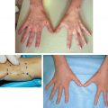
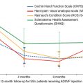 Chapter 20 SCLERODERMA AND FAT GRAFTING
 Chapter 16 IMPROVING SKIN QUALITY: AN EXPERIMENTAL AND CLINICAL STUDY OF THE EFFECTS OF FAT AND STROMAL CELLS ON THE SKIN
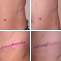
 Chapter 18 SCAR REMODELING WITH FAT GRAFTING AFTER BURN INJURY
 Chapter 21 TREATMENT OF DIFFICULT WOUNDS AND SCARS WITH FAT GRAFTS
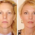 Chapter 23 SIMULTANEOUS FACELIFT AND FAT GRAFTING: COMBINED LIFTING AND FILLING FOR REJUVENATION OF THE AGING FACE
Stay updated, free articles. Join our Telegram channel

Full access? Get Clinical Tree


