Chapter 17 MICROFAT INJECTIONS WITH DISPOSABLE CANNULAS
Coleman’s method for grafting autologous adipose tissue is well established. The technique and its indications are solidly codified, with satisfactory results. 1 – 4 As early as 1992, Coleman had noticed that adipose tissue injections not only had a volumizing effect, but also that they improved skin quality. 5 , 6 With the appearance of microfat injections and the use of much smaller harvesting and injection equipment, the method has evolved over the past several years, and adipose tissue has become a valid subdermal filler. Advances in research have demonstrated the existence of stromal vascular fraction (SVF) in adipose tissue, which can be separated mechanically or by enzymatic digestion to obtain several tens of millions of cells containing between 3% and 5% stem cells. 7 , 8 In the near future, it will be possible to create blends of adipose tissue enriched with SVF, thereby improving trophic, angiogenic effects and giving real meaning to regenerative medicine and surgery.
Concepts and Rationales
Box 17-1 Comparison of Microfat Injection With Coleman Lipostructure
Coleman Lipostructure
A simple, validated technique with 20 years’ experience
No complications
Microfat Injection
Simpler technique
Validated through experimentation
More indications
The best subdermal filler product
Micrografting of autologous fatty tissue represents a major technologic evolution, thanks to the use of new disposable equipment, the characteristics of which were validated after years of experimentation. 9 With micrografting, it is now possible to harvest and graft adipose microparticles measuring approximately 0.5 mm and containing several hundred cells, and to get closer to deeper skin layers without the risk of causing surface irregularities.
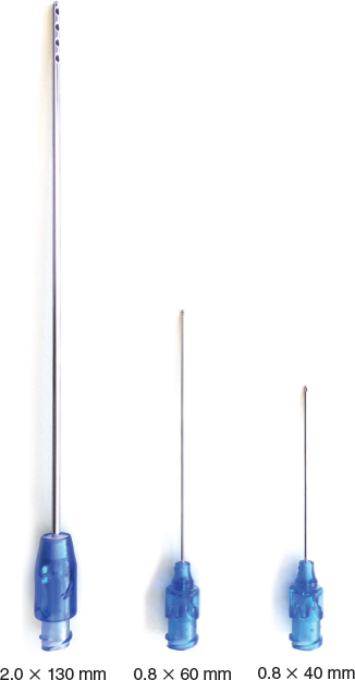
Micrografting techniques were made possible with the evolution of the equipment: harvesting cannulas are now smaller (14-gauge, 2 mm, 130 mm long), and grafting cannulas are as well (21-gauge, 0.8 mm, 40 or 60 mm long). Because of the small size of the cannulas, they are impossible to clean, so single-use cannulas are used.
Based on the work of Yoshimura and colleagues, 10 the “surviving zone” is less than 300 microns of the fat lobule. In this zone, both adipocytes and adipose-derived stromal cells (ADSCs) survive. This explains the very stable results with microinjection. Pallua and colleagues 11 have demonstrated that the viability and migration of isolated ADSCs, obtained with microharvested fat, were significantly higher and that microfat may be more suitable due to better cell viability for tissue engineering and regenerative surgery.
Harvested microfat may be mixed with platelet-rich plasma (PRP) in proportions of 10% to 50% and by counting the number of platelets to be injected using automated analyzers with impedance or fluorescence technique. Details about the percentage of PRP volume in the mixture and the number of platelets per volume of mixture should be systematically provided for a better understanding. If scarring appears to be a concern, this mixed product may be injected into the previously prepared lipoaspirate according to the concept of percutaneous aponeurotomy and lipofilling (PALF) developed by Khouri et al. 12
Indications
REPARATIVE AND RECONSTRUCTIVE SURGERY INDICATIONS
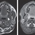
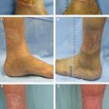
In reconstructive surgery, there are several indications for micrografting: for correction of adherent or depressed scars, atrophy after corticosteroid therapy, skin radiodermatitis, and in efforts to improve thinning skin, as in the hands. The technique also allows treatment of facial atrophy, particularly for scleroderma of the face. This technique is also effective for use inside and under burn scars.
Microfat injection is indicated for small volumes—in general, when less than 50 cc is required. In pediatric surgery, microfat injection is used to treat the sequelae of nasolabial and velopalatine clefts, to improve the volume and symmetry of the lips, and to address the posterior pharyngeal wall in patients with incompetence of the soft palate.
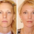
AESTHETIC INDICATIONS
Reduced facial volume and other signs of aging can be improved by replacing the missing tissue with fat to obtain a more youthful appearance. Microinjections are done at the cheekbones, temples, eyelids, nasolabial folds, marionette lines, lips, and chin.
The injection of fat into the subdermal plane will reduce facial wrinkles. This technique can be used to correct irregularities after rhinoplasty. Also, injection in the back of the hands and fingers have significant benefits for rejuvenating the appearance of the hands.
Related posts:
 Chapter 20 SCLERODERMA AND FAT GRAFTING
 Chapter 19 STRUCTURAL FAT GRAFTING FOR THE REGENERATION OF IRRADIATED TISSUE
 Chapter 16 IMPROVING SKIN QUALITY: AN EXPERIMENTAL AND CLINICAL STUDY OF THE EFFECTS OF FAT AND STROMAL CELLS ON THE SKIN
 Chapter 18 SCAR REMODELING WITH FAT GRAFTING AFTER BURN INJURY
 Chapter 21 TREATMENT OF DIFFICULT WOUNDS AND SCARS WITH FAT GRAFTS
 Chapter 23 SIMULTANEOUS FACELIFT AND FAT GRAFTING: COMBINED LIFTING AND FILLING FOR REJUVENATION OF THE AGING FACE
Stay updated, free articles. Join our Telegram channel

Full access? Get Clinical Tree


