Cervical Musculocutaneous Flap for Intraoral Lining
H. SAITO
EDITORIAL COMMENT
In effect, this is a variation of the platysmal myocutaneous flap. Because the blood supply to the platysma usually comes from branches along the facial artery, the flap must be designed in order not to jeopardize the blood supply.
INDICATIONS
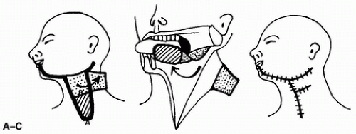
The lateral cervical musculocutaneous flap is used for reconstruction of lateral defects in the mouth and pharynx (Fig. 192.1). The median cervical musculocutaneous flap is used for reconstruction of the anterior median floor of the mouth (Fig. 192.2).
FLAP DESIGN AND DIMENSIONS
For the lateral cervical musculocutaneous flap, a vertical rectangular flap, usually 6 × 5 cm, is outlined on the lateral neck and is designed to be large enough to cover the defect. The tip can extend to just above the clavicle. The location of the base is usually 2 cm below the posterior half of the mandible and is 8 cm wide.
The procedure for the median cervical musculocutaneous flap is essentially the same as for the lateral flap, except the pedicle is based on the median submental region and is about 7 cm wide.
 FIGURE 192.1 Schematic drawing of the lateral cervical musculocutaneous flap. A: To make the subcutaneous tissue-platysma muscle pedicle of this flap, a laterally based epidermal-dermal flap was raised and turned back on itself (arrow). The thick line indicates the incision line of the vertical lateral cervical flap. B: The flap is rotated as the arrow indicates and sutured to the mucosal defect. C: Final external appearance. (From Saito et al., ref. 1, with permission.)
Stay updated, free articles. Join our Telegram channel
Full access? Get Clinical Tree
 Get Clinical Tree app for offline access

|


