Capillaritis (pigmented purpuric dermatoses)


Capillaritis (pigmented purpuric dermatoses)


Cord Sunderkötter and Thomas A. Luger


Evidence Levels:  A Double-blind study  B Clinical trial ≥ 20 subjects  C Clinical trial < 20 subjects  D Series ≥ 5 subjects  E Anecdotal case reports


image


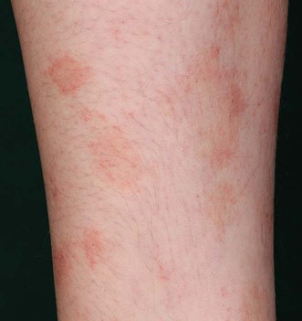
Capillaritis (a generic term for the various pigmented purpuric dermatoses) presents with the common feature of petechial macules or plaques and is characterized histologically by erythrocyte extravasation and perivascular infiltration with T lymphocytes. Lesions develop a characteristic brown to orange color due to hemosiderin deposits in macrophages. These conditions may also present with additional, distinctive, and sometimes overlapping, morphological patterns, which have given rise to several descriptive or eponymous names: papules in pigmented purpuric lichenoid dermatosis (of Gougerot and Blum) or in the rarely described granulomatous pigmented purpura (non-necrotizing granulomata with the concomitant lymphocytic infiltrate); eczematous spongiosis with pruritus in eczematoid-like purpura; annular forms with telangiectases and central clearing in purpura annularis telangiectodes (Majocchi disease); and often solitary, ochre–golden plaques or patches with band-like infiltrates including a grenz zone in lichen aureus. Sites of predilection are mostly both lower extremities. A form of unilateral linear capillaritis or involvement of other areas of the skin are rare.


The etiology of capillaritis, including these variants, is not known. The reason for extravasation of erythrocytes is not an inflammatory fibrinoid necrosis of vessels (no vasculitis). Possible pathophysiological factors that can be addressed therapeutically are a cell-mediated immune response, increased venous pressure, increased vascular permeability or vascular fragility due to subtle defects in the extracellular matrix.


Management strategy


Diagnostic hallmarks are the yellow–brown or orange patches with superimposed pinpoint cayenne pepper spots, which represent petechiae and persist with diascopy. The main differential diagnosis is leukocytoclastic or small vessel vasculitis. The discerning criterion is the lack of a palpable infiltrate in capillaritis, but there are variants of small vessel vasculitis with petechial maculae (e.g., as part of Sjögren syndrome). Thus, biopsies are warranted when in doubt.


In the differential diagnosis, allergic contact dermatitis may be hemorrhagic, mimicking capillaritis. Thrombocytopenia, hypergammaglobulinemic purpura of Waldenström, and the pigmented purpuric dermatitis-like variant of mycosis fungoides also need to be excluded. Although capillaritis has a benign course, it must be differentiated from these more serious diseases.


Usually there is no need for treatment unless the patient has pruritus or suffers from cosmetic disfigurement. Detection and avoidance of possible eliciting agents should always be attempted. Reported causes include:


When the etiology remains obscure therapy has to be empirical. However, immunohistochemical analyses have suggested a cell-mediated immune response, so treatment with local corticosteroids, calcineurin inhibitors, or psoralen plus UVA (PUVA) may be rational. Increased venous pressure (particularly in the legs) or exercise are not direct causes, but can aggravate capillaritis. In these cases compression stockings may be helpful.


There is some evidence for increased vascular permeability or vascular fragility due to subtle defects in the extracellular matrix. This may explain reported responses to bioflavonoids (which may be due to inhibition of elastase and hyaluronase and of leukocyte activation), ascorbic acid (antioxidant effects and perhaps reduction of vascular permeability), and calcium dobesilate (reduction of microvascular permeability in part by antioxidant properties).


Specific investigations


Stay updated, free articles. Join our Telegram channel

Aug 7, 2016 | Posted by in Dermatology | Comments Off on Capillaritis (pigmented purpuric dermatoses)

Full access? Get Clinical Tree

Get Clinical Tree app for offline access