Fig. 31.1
Classical bullous pemphigoid with tense bullae on the limb surrounded by erythema with infiltrated confluent urticarial plaques. Vesicles and eroded and crusted lesions are also observed
Fig. 31.2
Bullous pemphigoid presenting with widespread infiltrated urticarial and eczematous plaques
31.3 Non-bullous Forms and Unusual or Localized Variants
A number of patients are first evaluated and diagnosed with BP in the absence of any blistering. In a prospective Swiss cohort of 164 patients with BP, 80 % of patients showed localized or generalized bullous lesions together with other inflammatory skin lesions at time of diagnosis. Nevertheless, the remaining 20 % presented with no obvious blistering [23]. Another recent French study with 542 patients found identical figures with 21 % of the cases presenting with atypical features [22].
In these non-bullous forms, leading features of BP include excoriated, eczematous, papular, urticarial, or even only pigmented lesions associated with moderate to intractable pruritus that persists for weeks or even months (Figs. 31.3, 31.4, and 31.5). In this stage, diagnosis is particularly difficult and challenging. Several clinical variants of BP (Table 31.1) have been described with a variety of denominations, such as papular pemphigoid, prurigo and prurigo nodularis, erythema annulare centrifugum, erythroderma, ecthyma gangrenosum, and intertrigo-like and toxic epidermolysis-like lesions [11–29]. Localized bullous forms confined to paralyzed limbs and areas affected by radiotherapy, surgery, trauma, and burns as well as lesions limited around stomata, hemodialysis fistulae, the pretibial (Fig. 31.6) or umbilical area, the palmoplantar region mimicking dyshidrotic eczema (Fig. 31.7), and genital area have all been described anecdotally [30–41].
Fig. 31.3
Bullous pemphigoid associated with widespread annular and polycyclic lesions with infiltrated borders. There are scattered small vesicles and erosions
Fig. 31.4
Bullous pemphigoid with widespread eczematous lesions and excoriations
Fig. 31.5
Bullous pemphigoid presenting with isolated excoriated papules and linear postinflammatory hypopigmentation
Table 31.1
Spectrum of clinical presentations of bullous pemphigoid
Generalized lesions, trunk and limbs involved, variably accompanied by |
Vesicles and blisters |
Eczematous lesions |
Pigmented lesions |
Papular and/or urticarial lesions |
Erosive and crusted lesions |
Prurigo-like and prurigo nodularis-like lesions |
More uncommon presentations |
Ecthyma-like ulceration |
Erythema annulare centrifugum |
Erythema multiforme and toxic epidermolysis-like forms |
Erythrodermic type |
Localized variants |
Dyshidrosiform pemphigoid with isolated palmoplantar involvement |
Intertrigo with vegetating lesions |
Pretibial pemphigoid |
Peristomal pemphigoid |
Umbilical pemphigoid |
“Stump” pemphigoid on amputated limbs/extremity |
On paralyzed body sites |
On irradiated/traumatized body sites |
Brunsting-Perry form |
Localized lesions on the head and upper trunk |
Fig. 31.6
Prurigo nodularis-like variant of bullous pemphigoid with involvement of the lower limbs
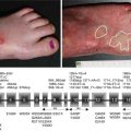
Fig. 31.7
Palm pompholyx-like involvement in bullous pemphigoid, which may occasionally represent the leading feature of the disease
Another peculiar rare variant of BP is lichen planus pemphigoides, which is associated with typical features of BP and lichen planus [42, 43]. Lichen planus pemphigoides usually occurs in the fifth decade, affects the limbs and extremities, and shows a relatively benign course. Finally, BP also occurs in children (“infantile” and “childhood” bullous pemphigoid) [44, 45]. Generalized involvement with also acral and facial distribution seems to be more frequently found in infantile bullous pemphigoid, whereas mucosal and genital (particularly in girls) involvement is observed in childhood pemphigoid in up to 44 % of cases [45].
As a matter of fact, all these different presentations represent different facets of the same disease. While the distinction of these forms is currently not justified based on the pathophysiology, they have at least the merit to help the clinicians to appreciate the various peculiar presentations. Nevertheless, it is time to rename BP, as was done in the case of mucous membrane pemphigoid. We propose replacing BP with the term cutaneous pemphigoid, a term with broader catchment in the spectrum of clinical BP presentations and with implications that the involvement of mucous membrane sites, while possible in up to 20 % of cases, is not predominant.
Continued careful clinical characterization of patients coupled with increased understanding of the pathophysiology including autoantibody specificity holds the promise of more precise diagnosis in the future and better understanding of the clinical phenotypes, just as patients with the inflammatory form of epidermolysis bullosa acquisita have clearly been distinguished from those with BP.
31.4 Mucosal Involvement
Mucosal lesions are observed in 10–30 % of patients [11–14]. In a recent prospective European cohort of BP patients, mucosal involvement, almost invariably of the oral mucosa, was found in 8 % of patients with BP [46]. In another prospective nationwide Swiss study, oral involvement was observed in up to 15 % of patients with again almost exclusively oral involvement [23]. The mucosae of the eyes, nose, pharynx, esophagus, and anogenital areas are rarely affected. In a recent French study encompassing 542 BP cases, 8 % had oral involvement without other mucosal sites affected [21].
31.5 Clinical Features and Clinical Diagnostic Criteria
Two French studies have provided strong evidence about the usefulness of certain clinical characteristics to make the diagnosis of BP [47, 48]. In patients having positive direct immunofluorescence studies with detectable linear IgG and/or C3 deposits along the dermoepidermal junction, a diagnosis of BP can be made with a high sensitivity and specificity (90 and 83 %, respectively) and a positive predictive value of 85 % when three of four clinical criteria are present: age greater than 70 years, absence of atrophic scars, absence of mucosal involvement, and absence of predominant bullous lesions on the neck and head. In contrast to later reports, in the Swiss cohort of BP patients, 20 and 15 % of the cases had neck/head involvement and mucosal lesions, respectively [23]. Future studies are needed to confirm whether the presence of neck and head involvement really represents a criterion that is reliable enough to dismiss the diagnosis of BP. Moreover, the fact that there is a subtype of BP which is typically localized on the head and neck, namely, the Brunsting-Perry form of BP, goes against this concept [49, 50].
31.6 Diagnostic Delays
Little data exist about the delay in diagnosis, that is, the interval between the development of the first symptoms and the time of definite diagnosis. Previous case reports described diagnostic delays varying between few weeks to more than one decade, but no systematic prospective analyses were available [18, 19, 47, 48, 51]. One early report described that in the presence of blisters, the diagnosis is made after about 6 weeks, whereas diagnosis may take years in the presence of nonspecific lesions [19, 51]. A French retrospective study assessing diagnostic delay in two large groups of patients reported a mean diagnostic delay of between 61 and 91 days, with two thirds of patients being diagnosed within 2 months [52]. In a multicenter European study about the immunological profile of 43 BP patients, the mean disease duration prior diagnosis was 3 months [46]. Finally, in another prospective survey, diagnosis of BP was made after a mean of 6.1 months and a median of 2.3 months after the development of the first symptoms. Specifically, while up to 70 % of the cases were diagnosed early within 4 months after the first symptoms, in the remaining cases, diagnosis was delayed up to several months [23]. The latter study showed that the more localized the disease at the first presentation is, the more difficult the diagnosis of BP. The diagnosis of BP seems to be more delayed in cases of lesions limited to one body area and in the case of head involvement, compared to patients with involvement of both the trunk and limb, independently of the presence of frank blistering.
31.7 Differential Diagnosis
Based on its multiple clinical facets, cutaneous pemphigoid is a clinical chameleon. In the various non-bullous presentations, it can bear close resemblance to a variety of dermatoses, including localized or generalized drug reactions, contact and allergic dermatitis, prurigo, fixed urticaria, urticarial vasculitis, arthropod reactions, scabies, ecthyma, or even pityriasis lichenoides. Detailed patient’s history, clinical evaluation, histopathologic features, and, above all, direct immunofluorescence microscopy studies are essential to distinguish these disorders from BP. Diseases of the pemphigus group can be easily differentiated on the basis of distinctive immunopathological features and the almost invariable presence of mucosal involvement (for the pemphigus vulgaris type) (see !Chaps. 23 and 24). In dermatitis herpetiformis, direct IF microscopy findings are distinctive (see Chap. 44). In contrast, the distinction of BP from epidermolysis bullosa acquisita or the so-called anti-p200 pemphigoid is often impossible based simply on clinical and histological features. Immunological testing (such as indirect immunofluorescence studies using split skin, specific ELISA, immunoblot studies) however can distinguish between these diseases (see Chap. 40). Paraneoplastic pemphigus, an autoimmune mucocutaneous disease associated with an underlying neoplasia and potential multiorgan involvement, might present with clinical features reminiscent of BP, although an invalidating stomatitis is almost invariably present. Immunopathological findings of PNP are distinctive enough to differentiate it from BP (see Chap. 25).
Related posts:
 Kindlin-1 and Its Role in Kindler Syndrome
 Cyclophosphamide in Autoimmune Blistering Diseases: Safety, Efficacy and Evidence Base
Management of Bullous Systemic Lupus Erythematosus
Cyclophosphamide in Autoimmune Blistering Diseases: Safety, Efficacy and Evidence Base
Management of Bullous Systemic Lupus Erythematosus
 Using Intravenous Immunoglobulins in Autoimmune Bullous Diseases
 Living with Epidermolysis Bullosa: Reviewing the Impact on Individuals’ Quality of Life
 Dermatitis Herpetiformis
Stay updated, free articles. Join our Telegram channel

Full access? Get Clinical Tree


