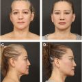
Male brow ptosis is a common condition that is challenging to treat. Multiple treatment options exist, but many traditional options result in either a significant scar or minimal improvement in brow position. Endoscopic brow lift in male patients is effective, well-supported in literature and should be considered for patients needing significant brow lift but wanting to avoid significant scarring.
Key points
- •
Treatment of male brow ptosis is challenging.
- •
There are multiple surgical methods for brow lifts, each with benefits and drawbacks.
- •
Endoscopic brow lift can result in an excellent lift with good longevity and should be considered for patients desiring effective brow lift and minimal visible scarring.
Introduction: nature of the problem
As patients age, one change they frequently experience is brow ptosis. Their symptoms include decreased superior visual fields, a tired appearance, and often headaches due to constant frontalis muscle activation. As the ptosis progresses, patients become increasingly bothered by the functional and cosmetic effects. Male patients generally have an especially prominent superior orbital rim and deep-set eyes, which can contribute to more significant visual field effects as brow ptosis progresses. Several surgical options for brow lift exist, demonstrating that no single option is perfect [ ].
Treatment of brow ptosis in male patients has proven to be especially difficult and somewhat controversial. The most effective methods of brow lifting often result in large scars, and those with minimal visible scarring often have little effect. Many surgeons avoid male brow lifts because they do not consider any of the options to be good.
Historically, the most effective brow lift has been a direct or mid-forehead lift. These result in excellent brow position, but generally leave a large scar not acceptable to a cosmetic patient. A pretrichial brow lift is often performed in female patients with good effect and the incision hidden in the hairline. In male patients, the dissection is often much more difficult, and the incision frequently remains visible, especially in patients with a receding hairline. A coronal brow lift is also effective but rarely performed because of the long dissection plane and resultant large scar [ , ].
Browpexy through a blepharoplasty incision (internal browpexy) or a small incision above the brow (external browpexy) results in small, less noticeable scars, but the result is brow stabilization more than true brow lifting [ , ]. Many surgeons perform blepharoplasty with transblepharoplasty browpexy for male patients with brow ptosis. In patients with significant brow ptosis, the minimal lift this provides is not sufficient.
Endoscopic-assisted brow lift was introduced in the 1990s. The endoscopic brow lift involves using three to five incisions, generally hidden in the hairline. Central dissection is carried out in either a pre-periosteal or, more commonly, a subperiosteal plane [ ]. Temporally, the dissection is carried out directly on the deep temporal fascia. The arcus marginalis and conjoint tendon are released, eyebrows are elevated, and fixation is performed, if needed, with sutures through screws or bone tunnels, Endotines, or tissue glue [ ]. An endoscope is often used for visualization, but in experienced hands is not required [ ].
The primary benefit of endoscopic brow lift is smaller incisions that are generally hidden in the hairline, improving cosmetic outcome. In addition, the surgical approach enables access to the corrugator and procerus muscles, which contribute to medial brow ptosis and prominent glabellar rhytids.
Shortly after its introduction, there was wide adoption of endoscopic brow lift. Many surgeons were pleased with the initial results but became dissatisfied with long-term (>2 year) results. As a result, many surgeons performed fewer endoscopic brow lifts or abandoned the procedure altogether [ ]. Most of the initial data regarding endoscopic brow lift were on female patients, with studies demonstrating that operations on males were a very small percentage of the total surgeries performed [ , ]. In addition, male pattern balding was considered a relative contraindication by many as some of the incisions would be visible [ ].
More recently, Jones and colleagues [ ] demonstrated in female patients that endoscopic brow lift resulted in excellent morphology and longevity of the brow lift with minimal relapse at the tail of the brow. Many surgeons have similar experience with their own patients and continue to perform endoscopic brow lifts.
Endoscopic brow lift has been well-documented for female patients, but less data are available in male patients. There are several studies which specifically study endoscopic brow lift in male patients [ ]. Overall, these demonstrated good eyebrow lift and minimal scarring, even in patients with male pattern balding. An additional study of both male and female patients did not give specific outcomes but noted good patient satisfaction in both groups [ ]. These studies all give robust evidence that endoscopic brow lift can be an effective tool for male brow lift with good patient satisfaction. Our experience also demonstrates excellent subjective results with objective measurements demonstrating sustained brow lift.
Surgical technique
Preoperative planning
Patients were selected for this procedure who had bothersome brow ptosis (often both superior visual field obstruction and cosmetic dissatisfaction), wanted brow lifting, and did not desire a large visible scar. They were counseled with a description of the procedure, postoperative care, and common side-effects, including forehead numbness. Many of these patients also had dermatochalasis or blepharoptosis and underwent blepharoplasty or blepharoptosis repair at the same time. Those meeting criteria underwent medical clearance with primary care to ensure they were medically optimized for surgery.
Prep and patient positioning
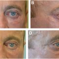
In the preoperative holding area, each patient was marked with one central, paired paramedian, and paired temporal marks ( Fig. 1 ). The central and paramedian marks were 1 cm long and generally oriented vertically, except in patients with receding hairlines, when the central incision was oriented horizontally to hide in a forehead rhytid. The temporal marks were 3 cm long and oriented horizontally. On each side, the expected location of the frontal branch of the facial nerve near the lateral canthus was marked, and the supraorbital and supratrochlear notches were palpated and marked. If the patient was undergoing concurrent blepharoplasty or ptosis repair, the eyelid markings could be made in either the preoperative area or in the operating room. The patient was placed in a supine position. For the brow lift portion of the procedure, the head was elevated (reverse Trendelenburg position) approximately 20°.

Related posts:
Stay updated, free articles. Join our Telegram channel

Full access? Get Clinical Tree


