There are an almost uncountable number of rashes that affect the trunk and extremities. This chapter reviews the most common causes of body rashes and includes discussions of atopic dermatitis, allergic contact dermatitis, nummular dermatitis, psoriasis, urticaria, scabies, bullous pemphigoid, pemphigus vulgaris, and more. atopic dermatitis allergic contact dermatitis nummular dermatitis psoriasis urticaria scabies bullous pemphigoid pemphigus vulgaris pityriasis rosea confluent and reticulated papillomatosis tinea corporis The American Academy of Dermatology (AAD) has developed atopic dermatitis (AD) criteria that are useful in evaluating patients for AD. The three essential criteria (pruritus, typical AD pattern, and chronic/relapsing course) should be used for screening patients with possible AD. If a patient does not display all three of these features, then they are very unlikely to have AD. Most adults with AD have had it since childhood, with many patients developing the condition during infancy. Some children present with a variant of AD called “follicular AD,” which is characterized by the development of itchy, follicular-based papules predominantly on the trunk. The differential diagnosis for AD is broad and is often confounded by the fact that AD frequently coexists with other conditions, predominantly allergic contact dermatitis (ACD) and irritant contact dermatitis (ICD). The differential diagnosis includes ACD, ICD, asteatotic or dry skin dermatitis, stasis dermatitis, cutaneous T cell lymphoma (CTCL) seborrheic dermatitis, and psoriasis. All patients with presumed AD should be approached systematically using the AAD criteria as previously discussed. A full-body skin examination should be performed in all patients to identify affected areas and to rule out signs of mimickers/concomitant skin conditions. Histopathologic examination is almost never necessary to confirm a diagnosis of AD; however, it can be important in ruling out mimickers, such as CTCL. Initial management of AD involves a four-pronged approach of dry skin care, topical antiinflammatory agents, trigger avoidance, and itch management. Dry skin care involves obtaining a list of all personal care products used and having an understanding of an individual’s bathing techniques (e.g., use of a luffa). First-line treatment for AD should include a medium-potency topical corticosteroid, such as triamcinolone 0.1% cream, applied twice daily for several weeks (with avoidance of application to facial skin or intertriginous areas to avoid skin thinning). Topical calcineurin inhibitors, such as tacrolimus or pimecrolimus 0.1%, can be used for maintenance therapy or for patients experiencing cutaneous adverse events from topical corticosteroid use. Identifying triggers in patients with AD can be very difficult but is necessary to avoid flares. There are now smartphone-based applications (colloquially known as “apps”) that can help patients track disease activity and input exposures to help identify possible triggers and predict possible flares. The most bothersome symptom of AD is intractable pruritus. Control of cutaneous disease correlates with itch control; however, itch-directed therapies are currently frequently ineffective. The biggest pitfall when managing AD in any location is failure to promote patient adherence to a dry skin care management regimen. Such a regimen is essential for achieving disease control and maintaining disease control in patients with AD; failure to implement a daily dry skin care regimen ensures treatment failure. Failure to identify coexisting dermatoses, such as ICD to soaps and ACD (including to topical corticosteroids), can interfere with successful AD management. Many patients with AD develop superinfections of their AD lesions because their skin barrier is impaired. The most common are impetigo and eczema herpeticum. Many patients or their parents request referral for food allergy testing because they want an explanation for why they or their child have developed AD. Blind elimination diets or use of immunoglobulin E (IgE) testing without confirmation with a double-blind food challenge is not recommended because it is rarely helpful. A diagnosis of CTCL should always be considered in patients with disease refractory to topicals, with new onset of disease in adulthood, with atypical-appearing patches/plaques, and/or with atypical distribution of patches/plaques. These patients require biopsies of untreated lesional skin. Specimens must be evaluated by a dermatopathologist given the difficulty of rendering a histologic diagnosis of CTCL. You have atopic dermatitis, also known as “eczema.” Atopic dermatitis is usually an inherited chronic skin condition (meaning there is no cure) that occurs in people who have skin that does not retain enough water and that does not adequately protect itself from outside allergens. Your skin is sensitive, prone to dryness, and susceptible to becoming itchy and developing rashes. Individuals with atopic dermatitis develop skin inflammation in response to the environment around them. This inflammation makes the skin red and itchy. Importantly, there is no one allergen that causes your skin disease. Additionally, dietary changes have not been reported in the medical literature to cure your disease. Treating your skin disease requires that you keep your skin healthy by moisturizing it and by avoiding exposing it to things that may irritate it. To keep your skin healthy, it is important that you have a special bathing regimen that promotes your skin health. When you shower, it is important that you use tepid water and that you limit your shower to less than 10 minutes. In the shower, you should use a gentle, unscented bar soap and should only apply soap to the armpits, groin, and areas that are visibly dirty. Do not use a luffa or a washcloth to apply soap because this may further irritate your skin. Pat, rather than rub, yourself dry with a towel. Immediately after drying off, you should apply a moisturizer to lock in the hydration that your skin received while you were showering. It is important that you not only hydrate your skin with a moisturizer after showering, but also that you moisturize at least twice a day. While your skin is still red and irritated, it is important that you do not apply any cosmetics or fragrances because these can inflame or irritate your skin. Your doctor has prescribed a topical steroid cream that you should use twice daily only for 7 to 10 days, at which time the redness and irritation should be gone. Do not apply this cream to your face, groin, underarms, or under your breasts unless told specifically to use the cream in these locations. Alternative steroid-sparing creams may also be prescribed for more chronic or intermittent use. These prescription creams are not a substitute for your moisturizer and should be used with your moisturizer, not instead of your moisturizer. Once the redness and irritation has resolved, you can stop the prescription cream, although you may want to restart it when the redness and irritation return. The topical steroid prescribed cream can cause side effects if you use it too frequently or if you use it for too long. These side effects include potential thinning of the skin, dilatation of blood vessels that can become more visible in the area of application, discoloration of the skin, and even an acne like rash. Continue your routine of twice-daily lubrication and shortened tepid bathing as part of your skin regimen. Atopic dermatitis is cyclical, meaning you will have times when your rash is much better and times when it is much worse. Different people flare for different reasons. You should try your best to identify things that are unique triggers for your atopic dermatitis. You will have to work with your doctor over time to find a skin care regimen that is right for you and that keeps your skin disease under control. Christian Gronbeck and Diane Whitaker-Worth ACD is a delayed-type hypersensitivity reaction to immunogenic agents that come into contact with the skin. Many patients who develop ACD have an impaired skin barrier, allowing small compounds to penetrate the skin and initiate an antigenic response. After initial allergen exposure, affected individuals progress through a sensitization phase, which involves the proliferation of antigen-specific T cells. After reexposure, activation of these T cells leads to an inflammatory response, which prompts more immediate cutaneous symptoms. Although the skin can be allergic to an innumerable number of compounds, the most common causes of ACD are nickel, fragrance mixtures, preservatives (e.g., formaldehyde), detergents, dyes (often in hair products), sunscreens (especially those containing benzophenones), topical medications (e.g., minoxidil, neomycin, bacitracin), acrylates, and urushiol (a chemical in poison ivy, poison sumac, and poison oak). Although ACD occurs equally among all races, it is more commonly seen in female patients, likely because of the higher prevalence of piercings and the increased number of personal care products used by women. The differential for ACD includes AD, ICD, psoriasis, and seborrheic dermatitis. A total-body skin examination and patient history is necessary to support the diagnosis of ACD. Patch testing is the gold standard in confirming the diagnosis of ACD and identifying likely allergens: Patients should be counseled to avoid identified or highly suspected allergens. Patients with chronic ACD may require more extensive management that includes oral immunomodulatory drugs (e.g., methotrexate, mycophenolate) or ultraviolet (UV) light treatment. Nevertheless, these patients are typically best referred to a dermatologist for close management and to ensure their allergens have been adequately identified through patch testing. If there is a partial but inadequate response after a 4-week trial of topical corticosteroid monotherapy: Allergic contact dermatitis is a skin rash caused by a skin allergy to certain products, chemicals, or medications in your day-to-day environment. Common allergens include shampoos, soaps, nickel (on your watch or pants button), fragrances, cosmetic agents, creams, and some plants, such as poison ivy. Importantly, this type of allergy is not life-threatening. We generally manage this rash in a few ways. First, we want to identify what you are allergic to. To determine this, we will refer you to a dermatologist for patch testing. Patch testing is a painless procedure in which small pieces of potential allergens will be taped to your back and left in place for 2 days. If we identify what you are allergic to, we recommend that you avoid that allergen to see if your symptoms resolve. We also have treatments available that can help to resolve your skin rash while we are waiting to determine what you are allergic to. We have prescribed you a topical corticosteroid cream, which you should apply to the rash twice daily. The most common side effect people notice from this cream is that it can cause the skin to thin slightly and the blood vessels to become more noticeable in the surrounding area. Most patients see their rash begin to resolve within a few weeks of consistently using the cream. In addition to using these treatments, you may also find it helpful to hydrate your skin with moisturizers. Christian Gronbeck and Diane Whitaker-Worth Nummular dermatitis (eczema) is an inflammatory skin condition that derives its name from its characteristic coin-shaped lesions (Fig. 3.4). Nummular dermatitis is likely a clinical variant of atopic eczema and ACD because it shares features of both conditions.
3: Body dermatitis
Abstract:
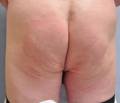
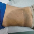
Atopic dermatitis—trunk and extremities
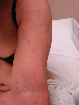
Clinical features
Differential diagnosis
Work-up
Initial steps in management
Dry skin care
Topical antiinflammatory agents
Trigger avoidance
Itch management
Other therapies
Warning signs/common pitfalls
Counseling
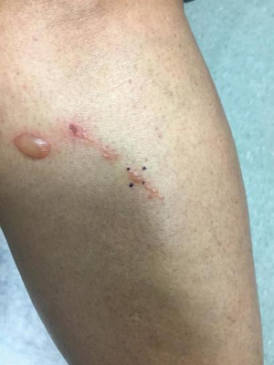
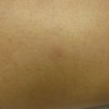
Allergic contact dermatitis—trunk
Clinical features
Course of development
Initial exposure
Subsequent exposures
Chronic allergic contact dermatitis
Common clinical presentations
Differential diagnosis
Work-up
Initial steps in management
General management comments
Allergen avoidance and protection
Minimization of existing skin inflammation
Management of chronic allergic contact dermatitis
Partial but inadequate response
Warning signs/common pitfalls
Counseling
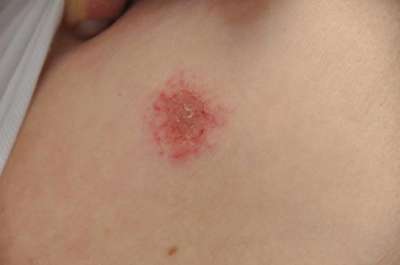
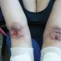
Nummular dermatitis
Clinical features
![]()
Stay updated, free articles. Join our Telegram channel

Full access? Get Clinical Tree


Plastic Surgery Key
Fastest Plastic Surgery & Dermatology Insight Engine