Benign neoplasms of the skin are commonly seen by all physicians. It is vital to distinguish these proliferations from malignant lesions. Despite their benign nature, many of these neoplasms cause aesthetic or symptomatic distress and require removal. Some of these benign neoplasms may serve as markers for internal disease or genetic syndromes. Multiple forms of therapy, including excision, cryotherapy, curettage, laser therapy, and pharmacotherapy, are available. This article discusses the epidemiology, clinical presentation, and recent advances in the management of these benign lesions.
Key point
- •
It is essentially to rule out malignant melanoma before trying to remove nevi Propanolol is a safe and effective treatment for infantile hemangiomas Benign cysts and lipomas are generally best treated with surgical exsion.
Disorders of melanocytes
- 1.
It is vital, especially in patients with multiple nevi, to rule out malignant melanoma, especially before removal with CO 2 , Er:YAG, and Nd:YAG lasers.
- 2.
Blue nevi can be misdiagnosed as melanoma, but dermatoscopic results of homogeneous pigmentation support the diagnosis of blue nevi.
- 3.
In patients with multiple halo nevi, screening for vitiligo is essential, especially in patients with family history of autoimmune disorders.
Acquired Nevi (Moles)
Acquired nevi are one of the most frequently acquired new growths in white patients, with average adults accumulating, on average, 20 nevi. They tend to appear in early childhood and reach a maximum in young adulthood. One recent study noted that 83% of newborns examined within 48 hours of birth presented with nevi. They undergo gradual fibrosis, with most disappearing by age 60. African Americans and Asians tend to less frequently present with nevi; however, when they do, the nevi are more likely to occur on palms, soles, and nail beds. Sun exposure and ultraviolet radiation have been identified throughout literature as the major factors that induce nevi on exposed surfaces.
Benign nevi are common, small (<1 cm diameter), circumscribed, pigmented macules, papules, or nodules. They tend to be asymptomatic with uniform shape and color, ranging from pink to black. Nevi are composed of nests of nevus cells located in the epidermis, dermis, and/or (rarely) subcutaneous tissue, with the following progression.
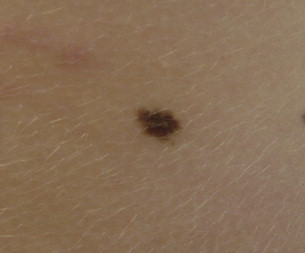
Junctional melanocytic nevi
Defined as nevus cell nests on the epidermal side of the basement membrane, making them intraepidermal structures ( Fig. 1 ), these tend to be flat dark nevi because the melanocytes have the greatest capacity to form melanin in this layer.
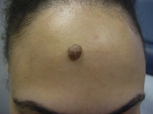
Compound melanocytic nevi
Defined as nevus cell nests invading the papillary dermis, making these lesions intraepidermal and dermal ( Fig. 2 ), these present as raised lesions of variable color.
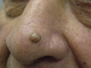
Dermal melanocytic nevi
Defined as nevus cells located exclusively in the dermis, due to melanocytes losing their capacity to make melanin as they penetrate into the dermis, these present as elevated, pink to light brown lesions ( Fig. 3 ).
Although nevi are benign findings individually, the risk of melanoma is related to the number and size of the nevi. Physicians must be aware of the dysplastic nevus syndrome in which dysplastic nevi are precursors to malignant melanoma. Treatment, although not necessary, usually involves surgical excision. Recently, it has been reported that, although traditionally CO 2 or Er:YAG lasers have been used to remove melanocytic nevi, 1064 nm Q-switched Nd:YAG lasers also provide a safe and effective treatment modality.
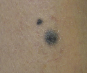
Blue Nevi
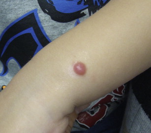
Blue nevi appear as small, sharply defined, round, dark blue, or grayish-blue lesions that most commonly develop during childhood ( Fig. 4 ). These tend to be slow growing nodules covered by smooth, intact epidermis. These lesions are composed of melanocytes limited to the dermis, with an intimate association with the fibroblasts of the dermis. Blue nevi may occur anywhere on the body, but most commonly on the face, neck, hands, and arms. Unfortunately, because they can be acquired lesions, they can frequently be misdiagnosed as melanoma or cutaneous metastasis of melanoma. Therefore, when melanoma is suspected, surgical excision is mandatory.
Halo Nevus
Halo nevi appear as nevi encircled by an oval to round halo of sharply demarcated leukoderma or depigmentation. The leukoderma is based on the decrease of melanin in melanocytes and/or disappearance of melanocytes at the dermal-epidermal junction. These halo nevi can occur around blue nevi, congenital nevomelanocytic nevi, Spitz nevi, primary melanoma, melanoma metastases, dermatofibroma, or neurofibroma. Overall prevalence is 1%, seen with spontaneous onset within the first three decades of life, generally with multiple lesions on the back.
Many recent studies have identified that those patients with multiple halo nevi may be more likely to develop vitiligo, especially in those with family history of autoimmune disorders. This lesion is considered an autoimmune response, mediated by T lymphocytes and, therefore, has been associated with other autoimmune conditions, including vitiligo, celiac disease, atopic dermatitis, alopecia areata, and Hashimoto thyroiditis.
Spitz Nevus
Spitz Nevi can occur across all ages, though typically they occur in patients less than 10 years old ( Fig. 5 ). The lesions arise rapidly, usually over several months, and are distributed most frequently on the head and neck (37%) and lower extremities (28%), with less than 6% located on the back.
The lesions are solitary, well-defined, dome-shaped, hairless, firm, small (<1 cm) papules or nodules. They are typically pink to red due to the limited melanin content and increased vascularity within these nevi. About 10% may be pigmented, however, ranging in color from tan to brown to black. Nevi that are greater than 1 cm diameter, present with asymmetry, and/or irregular borders, shape, and color, should be examined more closely for potential malignancy.
Spitz tumors have long been histologically challenging diagnoses; however, histologic examination must be done to confirm the clinical diagnosis of Spitz nevi. Evidence of neat organizational attributes, such as symmetry, maturation with descent, distinct margins, and small size are typical. Epidermal hyperplasia is common with melanocytic nests weaving neatly between keratinocytes (unlike melanoma, which tends to be more disordered). Furthermore, Kamino bodies (eosinophilic aggregates) may be present intraepidermally and at the dermoepidermal junction. Histopathologic features that suggest more aggressive behavior are increased mitotic activity, mitoses near the base of the lesion, ulceration, deeper extension, and inflammation.
Rare instances of metastasis have been observed with Spitz tumors. Atypical Spitz tumors greater than 1 cm diameter and ulcerated, with subcutaneous involvement and mitotic figures, are more likely to show micrometastasis. Adult Spitz nevi are usually excised, whereas pediatric cases are sometimes managed nonsurgically due to the lower probability of true melanoma. When these nevi are removed, it is advised that the lesion be completely excised due to a high frequency of recurrence. Due to this risk, it is advisable also to re-excise a Spitz nevus if the margins are involved. Sentinel node biopsy has been suggested as a possible diagnostic technique, but it has not been met with favor as a global screening tool. Up to 40% of Spitz nevi cases were associated with regional metastasis, which was associated with an extremely low rate of mortality.
Nevus Sebaceous
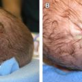
Nevus sebaceous presents at birth or within the first few months of life, with equal incidence in males and females. This hamartoma most commonly presents as a solitary papillomatous yellow-orange linear or oval plaque on the scalp or face. Lesions on the scalp are associated with alopecia. This lesion has been seen in association with the SCALP syndrome, which describes a constellation of sebaceous nevus with associated central nervous system malformations, aplasia cutis congenita, limbal dermoid, and pigmented nevi.
These lesions, similar to other types of nevi, progress through several stages. During the first few months after birth there are an increased number of sebaceous glands with diminution in hair follicles due to hormonal influences. During the next phase, there is a slight papillomatous epithelial hyperplasia and increased number of underdeveloped sebaceous glands, eccrine glands, and miniature hairs. Then rapid growth occurs at puberty with enlarged sebaceous glands, apocrine gland maturation, verrucous hyperplasia of the epidermis, and decreased number of normal hair follicles.
Benign tumors, including syringocystadenoma papilliferum, trichoblastoma, and trichilemmoma, have been reported developing secondarily within nevus sebaceous at an incidence of 16%. Malignant tumors such as basal cell carcinoma were reported at an incidence of 8%. There has been recent controversy about the utility of prophylactic excision of these nevi to avoid development of such malignancies and, therefore, further studies must be performed before a recommendation is made. Definitive treatment would involve surgical excision with narrow margins. In many cases, patients also desire removal secondary to cosmetic concerns because these nevi tend to occur on the face or cause alopecia when on the scalp.
Disorders of melanocytes
- 1.
It is vital, especially in patients with multiple nevi, to rule out malignant melanoma, especially before removal with CO 2 , Er:YAG, and Nd:YAG lasers.
- 2.
Blue nevi can be misdiagnosed as melanoma, but dermatoscopic results of homogeneous pigmentation support the diagnosis of blue nevi.
- 3.
In patients with multiple halo nevi, screening for vitiligo is essential, especially in patients with family history of autoimmune disorders.
Acquired Nevi (Moles)
Acquired nevi are one of the most frequently acquired new growths in white patients, with average adults accumulating, on average, 20 nevi. They tend to appear in early childhood and reach a maximum in young adulthood. One recent study noted that 83% of newborns examined within 48 hours of birth presented with nevi. They undergo gradual fibrosis, with most disappearing by age 60. African Americans and Asians tend to less frequently present with nevi; however, when they do, the nevi are more likely to occur on palms, soles, and nail beds. Sun exposure and ultraviolet radiation have been identified throughout literature as the major factors that induce nevi on exposed surfaces.
Benign nevi are common, small (<1 cm diameter), circumscribed, pigmented macules, papules, or nodules. They tend to be asymptomatic with uniform shape and color, ranging from pink to black. Nevi are composed of nests of nevus cells located in the epidermis, dermis, and/or (rarely) subcutaneous tissue, with the following progression.
Junctional melanocytic nevi
Defined as nevus cell nests on the epidermal side of the basement membrane, making them intraepidermal structures ( Fig. 1 ), these tend to be flat dark nevi because the melanocytes have the greatest capacity to form melanin in this layer.
Compound melanocytic nevi
Defined as nevus cell nests invading the papillary dermis, making these lesions intraepidermal and dermal ( Fig. 2 ), these present as raised lesions of variable color.
Dermal melanocytic nevi
Defined as nevus cells located exclusively in the dermis, due to melanocytes losing their capacity to make melanin as they penetrate into the dermis, these present as elevated, pink to light brown lesions ( Fig. 3 ).
Although nevi are benign findings individually, the risk of melanoma is related to the number and size of the nevi. Physicians must be aware of the dysplastic nevus syndrome in which dysplastic nevi are precursors to malignant melanoma. Treatment, although not necessary, usually involves surgical excision. Recently, it has been reported that, although traditionally CO 2 or Er:YAG lasers have been used to remove melanocytic nevi, 1064 nm Q-switched Nd:YAG lasers also provide a safe and effective treatment modality.
Blue Nevi
Blue nevi appear as small, sharply defined, round, dark blue, or grayish-blue lesions that most commonly develop during childhood ( Fig. 4 ). These tend to be slow growing nodules covered by smooth, intact epidermis. These lesions are composed of melanocytes limited to the dermis, with an intimate association with the fibroblasts of the dermis. Blue nevi may occur anywhere on the body, but most commonly on the face, neck, hands, and arms. Unfortunately, because they can be acquired lesions, they can frequently be misdiagnosed as melanoma or cutaneous metastasis of melanoma. Therefore, when melanoma is suspected, surgical excision is mandatory.
Halo Nevus
Halo nevi appear as nevi encircled by an oval to round halo of sharply demarcated leukoderma or depigmentation. The leukoderma is based on the decrease of melanin in melanocytes and/or disappearance of melanocytes at the dermal-epidermal junction. These halo nevi can occur around blue nevi, congenital nevomelanocytic nevi, Spitz nevi, primary melanoma, melanoma metastases, dermatofibroma, or neurofibroma. Overall prevalence is 1%, seen with spontaneous onset within the first three decades of life, generally with multiple lesions on the back.
Many recent studies have identified that those patients with multiple halo nevi may be more likely to develop vitiligo, especially in those with family history of autoimmune disorders. This lesion is considered an autoimmune response, mediated by T lymphocytes and, therefore, has been associated with other autoimmune conditions, including vitiligo, celiac disease, atopic dermatitis, alopecia areata, and Hashimoto thyroiditis.
Spitz Nevus
Spitz Nevi can occur across all ages, though typically they occur in patients less than 10 years old ( Fig. 5 ). The lesions arise rapidly, usually over several months, and are distributed most frequently on the head and neck (37%) and lower extremities (28%), with less than 6% located on the back.
The lesions are solitary, well-defined, dome-shaped, hairless, firm, small (<1 cm) papules or nodules. They are typically pink to red due to the limited melanin content and increased vascularity within these nevi. About 10% may be pigmented, however, ranging in color from tan to brown to black. Nevi that are greater than 1 cm diameter, present with asymmetry, and/or irregular borders, shape, and color, should be examined more closely for potential malignancy.
Spitz tumors have long been histologically challenging diagnoses; however, histologic examination must be done to confirm the clinical diagnosis of Spitz nevi. Evidence of neat organizational attributes, such as symmetry, maturation with descent, distinct margins, and small size are typical. Epidermal hyperplasia is common with melanocytic nests weaving neatly between keratinocytes (unlike melanoma, which tends to be more disordered). Furthermore, Kamino bodies (eosinophilic aggregates) may be present intraepidermally and at the dermoepidermal junction. Histopathologic features that suggest more aggressive behavior are increased mitotic activity, mitoses near the base of the lesion, ulceration, deeper extension, and inflammation.
Rare instances of metastasis have been observed with Spitz tumors. Atypical Spitz tumors greater than 1 cm diameter and ulcerated, with subcutaneous involvement and mitotic figures, are more likely to show micrometastasis. Adult Spitz nevi are usually excised, whereas pediatric cases are sometimes managed nonsurgically due to the lower probability of true melanoma. When these nevi are removed, it is advised that the lesion be completely excised due to a high frequency of recurrence. Due to this risk, it is advisable also to re-excise a Spitz nevus if the margins are involved. Sentinel node biopsy has been suggested as a possible diagnostic technique, but it has not been met with favor as a global screening tool. Up to 40% of Spitz nevi cases were associated with regional metastasis, which was associated with an extremely low rate of mortality.
Nevus Sebaceous
Nevus sebaceous presents at birth or within the first few months of life, with equal incidence in males and females. This hamartoma most commonly presents as a solitary papillomatous yellow-orange linear or oval plaque on the scalp or face. Lesions on the scalp are associated with alopecia. This lesion has been seen in association with the SCALP syndrome, which describes a constellation of sebaceous nevus with associated central nervous system malformations, aplasia cutis congenita, limbal dermoid, and pigmented nevi.
These lesions, similar to other types of nevi, progress through several stages. During the first few months after birth there are an increased number of sebaceous glands with diminution in hair follicles due to hormonal influences. During the next phase, there is a slight papillomatous epithelial hyperplasia and increased number of underdeveloped sebaceous glands, eccrine glands, and miniature hairs. Then rapid growth occurs at puberty with enlarged sebaceous glands, apocrine gland maturation, verrucous hyperplasia of the epidermis, and decreased number of normal hair follicles.
Benign tumors, including syringocystadenoma papilliferum, trichoblastoma, and trichilemmoma, have been reported developing secondarily within nevus sebaceous at an incidence of 16%. Malignant tumors such as basal cell carcinoma were reported at an incidence of 8%. There has been recent controversy about the utility of prophylactic excision of these nevi to avoid development of such malignancies and, therefore, further studies must be performed before a recommendation is made. Definitive treatment would involve surgical excision with narrow margins. In many cases, patients also desire removal secondary to cosmetic concerns because these nevi tend to occur on the face or cause alopecia when on the scalp.
Related posts:

Stay updated, free articles. Join our Telegram channel

Full access? Get Clinical Tree


